Graves Amanda B, Morse Robert P, Chao Alex, Iniguez Angelina, Goulding Celia W, Liptak Matthew D
Department of Chemistry, University of Vermont , Burlington, Vermont 05405, United States.
Inorg Chem. 2014 Jun 16;53(12):5931-40. doi: 10.1021/ic500033b. Epub 2014 Jun 5.
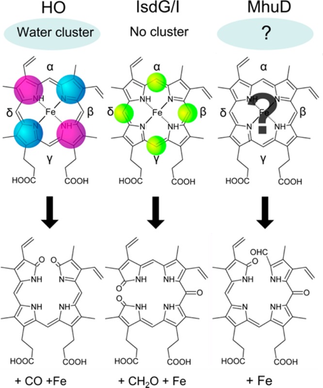
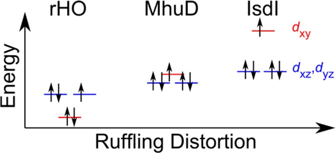
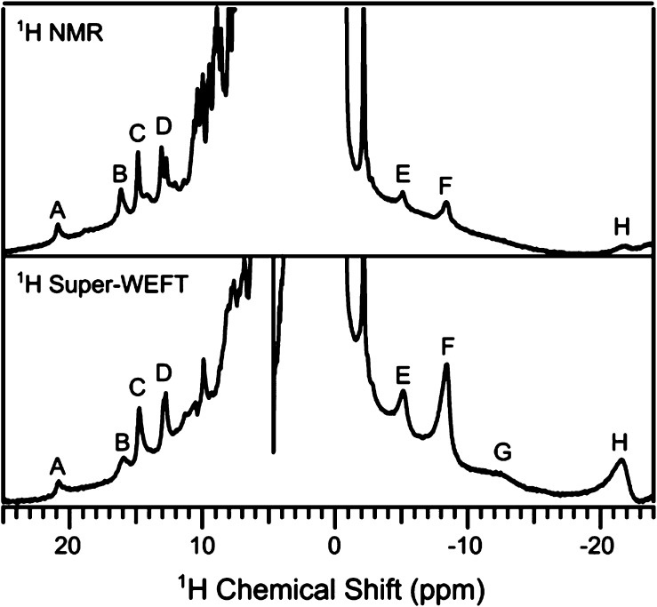
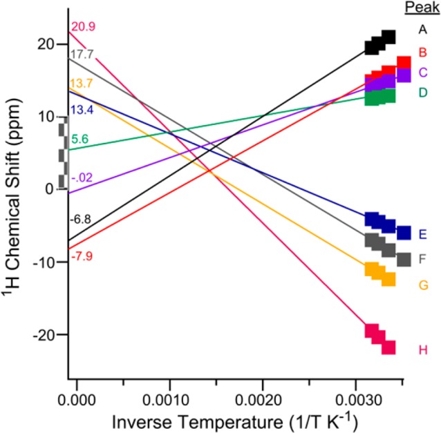
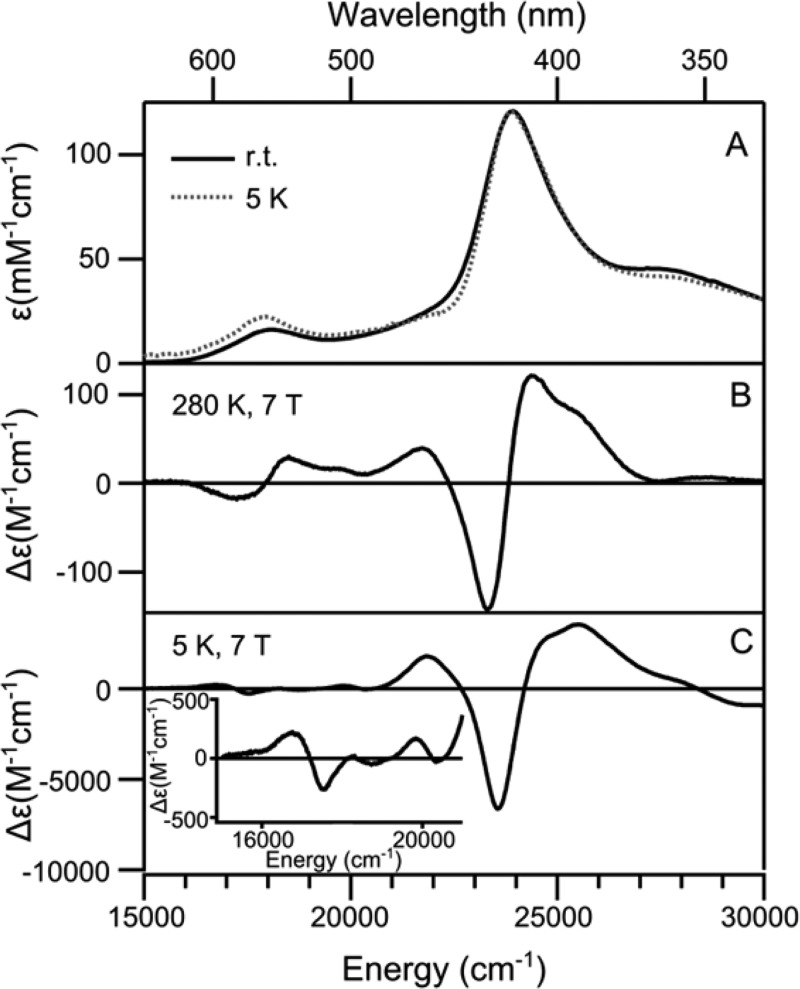
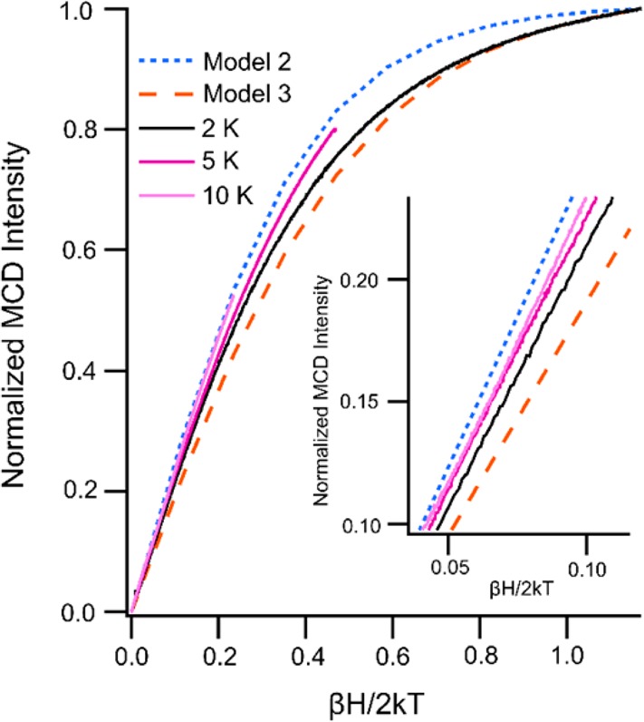
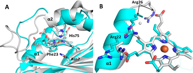
Mycobacterium heme utilization degrader (MhuD) is a heme-degrading protein from Mycobacterium tuberculosis responsible for extracting the essential nutrient iron from host-derived heme. MhuD has been previously shown to produce unique organic products compared to those of canonical heme oxygenases (HOs) as well as those of the IsdG/I heme-degrading enzymes from Staphylococcus aureus. Here, we report the X-ray crystal structure of cyanide-inhibited MhuD (MhuD-heme-CN) as well as detailed (1)H nuclear magnetic resonance (NMR), UV/vis absorption, and magnetic circular dichroism (MCD) spectroscopic characterization of this species. There is no evidence for an ordered network of water molecules on the distal side of the heme substrate in the X-ray crystal structure, as was previously reported for canonical HOs. The degree of heme ruffling in the crystal structure of MhuD is greater than that observed for HO and less than that observed for IsdI. As a consequence, the Fe 3dxz-, 3dyz-, and 3dxy-based MOs are very close in energy, and the room-temperature (1)H NMR spectrum of MhuD-heme-CN is consistent with population of both a (2)Eg electronic state with a (dxy)(2)(dxz,dyz)(3) electron configuration, similar to the ground state of canonical HOs, and a (2)B2g state with a (dxz,dyz)(4)(dxy)(1) electron configuration, similar to the ground state of cyanide-inhibited IsdI. Variable temperature, variable field MCD saturation magnetization data establishes that MhuD-heme-CN has a (2)B2g electronic ground state with a low-lying (2)Eg excited state. Our crystallographic and spectroscopic data suggest that there are both structural and electronic contributions to the α-meso regioselectivity of MhuD-catalyzed heme cleavage. The structural distortion of the heme substrate observed in the X-ray crystal structure of MhuD-heme-CN is likely to favor cleavage at the α- and γ-meso carbons, whereas the spin density distribution may favor selective oxygenation of the α-meso carbon.
结核分枝杆菌血红素利用降解酶(MhuD)是一种来自结核分枝杆菌的血红素降解蛋白,负责从宿主来源的血红素中提取必需营养铁。与典型的血红素加氧酶(HOs)以及金黄色葡萄球菌的IsdG/I血红素降解酶相比,MhuD先前已被证明能产生独特的有机产物。在此,我们报告了氰化物抑制的MhuD(MhuD-血红素-CN)的X射线晶体结构,以及该物种详细的氢核磁共振(NMR)、紫外/可见吸收和磁圆二色性(MCD)光谱表征。在X射线晶体结构中,没有证据表明在血红素底物远端存在有序的水分子网络,这与之前报道的典型HOs情况不同。MhuD晶体结构中血红素的褶皱程度大于HO观察到的情况,小于IsdI观察到的情况。因此,基于Fe 3dxz、3dyz和3dxy的分子轨道能量非常接近,MhuD-血红素-CN的室温氢核磁共振谱与具有(dxy)(2)(dxz,dyz)(3)电子构型的(2)Eg电子态以及具有(dxz,dyz)(4)(dxy)(1)电子构型的(2)B2g态的布居情况一致,前者类似于典型HOs的基态,后者类似于氰化物抑制的IsdI的基态。变温、变场MCD饱和磁化数据表明,MhuD-血红素-CN具有(2)B2g电子基态和低能级的(2)Eg激发态。我们的晶体学和光谱数据表明,MhuD催化的血红素裂解的α-中位区域选择性存在结构和电子方面的贡献。在MhuD-血红素-CN的X射线晶体结构中观察到的血红素底物的结构畸变可能有利于在α-和γ-中位碳处裂解,而自旋密度分布可能有利于α-中位碳的选择性氧化。