Zhu Huaqi, Sun Qiman, Tan Changjun, Xu Min, Dai Zhi, Wang Zheng, Fan Jia, Zhou Jian
Liver Cancer Institute, Zhongshan Hospital, Fudan University, Shanghai 200032, P.R. China.
Mol Med Rep. 2014 Aug;10(2):585-92. doi: 10.3892/mmr.2014.2302. Epub 2014 Jun 5.
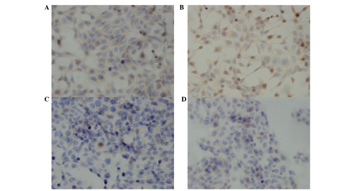
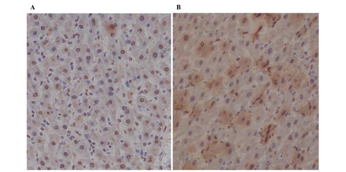
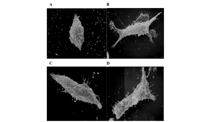
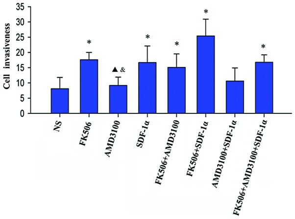
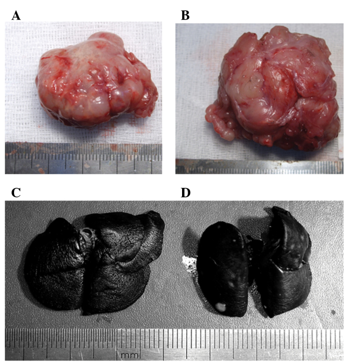
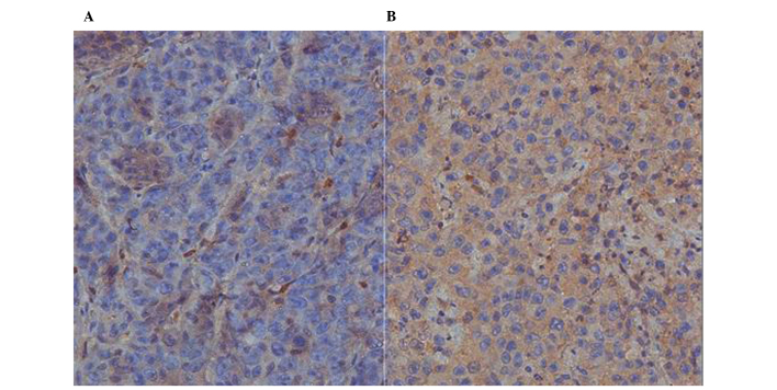
The aim of our study was to elucidate the effect of tacrolimus (FK506) and of C-X-C chemokine receptor type 4 (CXCR4), which is a receptor specific to the stromal cell-derived factor-1α (SDF‑1α), on growth and metastasis of hepatocellular carcinoma (HCC). Following treatment with different concentrations of FK506, AMD3100 or normal saline (NS), the proliferation of Morris rat hepatoma 3924A (MH3924A) cells was measured by the MTT assay, the expression of CXCR4 was analyzed with immunohistochemistry, and the morphological changes and the invasiveness of cells were studied with a transwell assay and under a scanning electron microscope, respectively. In addition, August Copenhagen Irish rat models implanted with tumor were used to examine the pathological changes and invasiveness of tumor in vivo, the expression of CXCR4 in tumor tissues and the expression of SDF‑1α in the adjacent tissues to the HCC ones, using immunohistochemistry. In vitro, FK506 (100‑1,000 µg/l) significantly promoted the proliferation of MH3924A cells (P<0.01), and increased the expression of CXCR4 in MH3924A cells, albeit with no significance (P>0.05). By contrast, AMD3100 had no effect on the proliferation of MH3924A cells, but significantly reduced the expression of CXCR4 (P<0.05). The invasiveness of MH3924A cells was significantly (P<0.01) enhanced following treatment with FK506, SDF‑1α, FK506 + AMD3100, FK506 + SDF‑1α or FK506 + AMD3100 + SDF‑1α. In vivo, tumor weight (P=0.041), lymph node metastasis (P=0.002), the number of pulmonary nodules (P=0.012), the expression of CXCR4 in tumor tissues (P=0.048) and that of SDF‑1α in adjacent tissues (P=0.026) were significantly different between the FK506-treated and the NS group. Our results suggest that FK506 promotes the proliferation of MH3924A cells and the expression of CXCR4 and SDF‑1α in vivo. Therefore, inhibiting the formation of the CXCR4/SDF‑1α complex may partly reduce the promoting effect of FK506 on HCC.
我们研究的目的是阐明他克莫司(FK506)以及基质细胞衍生因子-1α(SDF-1α)的特异性受体C-X-C趋化因子受体4(CXCR4)对肝细胞癌(HCC)生长和转移的影响。用不同浓度的FK506、AMD3100或生理盐水(NS)处理后,采用MTT法检测Morris大鼠肝癌3924A(MH3924A)细胞的增殖情况,用免疫组织化学法分析CXCR4的表达,分别用Transwell实验和扫描电子显微镜研究细胞的形态变化和侵袭能力。此外,将荷瘤的奥古斯塔·哥本哈根·爱尔兰大鼠模型用于检测体内肿瘤的病理变化和侵袭能力,并用免疫组织化学法检测肿瘤组织中CXCR4的表达以及肝癌相邻组织中SDF-1α的表达。在体外,FK506(100-1000μg/L)显著促进MH3924A细胞的增殖(P<0.01),并增加MH3924A细胞中CXCR4的表达,尽管差异无统计学意义(P>0.05)。相比之下,AMD3100对MH3924A细胞的增殖无影响,但显著降低CXCR4的表达(P<0.05)。用FK506、SDF-1α、FK506+AMD3100、FK506+SDF-1α或FK506+AMD3100+SDF-1α处理后,MH3924A细胞的侵袭能力显著增强(P<0.01)。在体内,FK506处理组与NS组相比,肿瘤重量(P=0.041)、淋巴结转移(P=0.002)、肺结节数量(P=0.012)、肿瘤组织中CXCR4的表达(P=0.048)以及相邻组织中SDF-1α的表达(P=0.026)均有显著差异。我们的结果表明,FK506促进MH3924A细胞的增殖以及体内CXCR4和SDF-1α的表达。因此,抑制CXCR4/SDF-1α复合物的形成可能部分降低FK506对肝癌的促进作用。