Zhou Hao, Yang Junjie, Xin Ting, Zhang Tao, Hu Shunyin, Zhou Shanshan, Chen Guanghui, Chen Yundai
Department of Cardiology, Chinese People's Liberty Army General Hospital, Beijing 100853, P.R. China.
Department of Cardiology, Tianjin First Central Hospital, Tianjin 300192, P.R. China.
Mol Med Rep. 2015 Jun;11(6):4063-72. doi: 10.3892/mmr.2015.3243. Epub 2015 Jan 22.
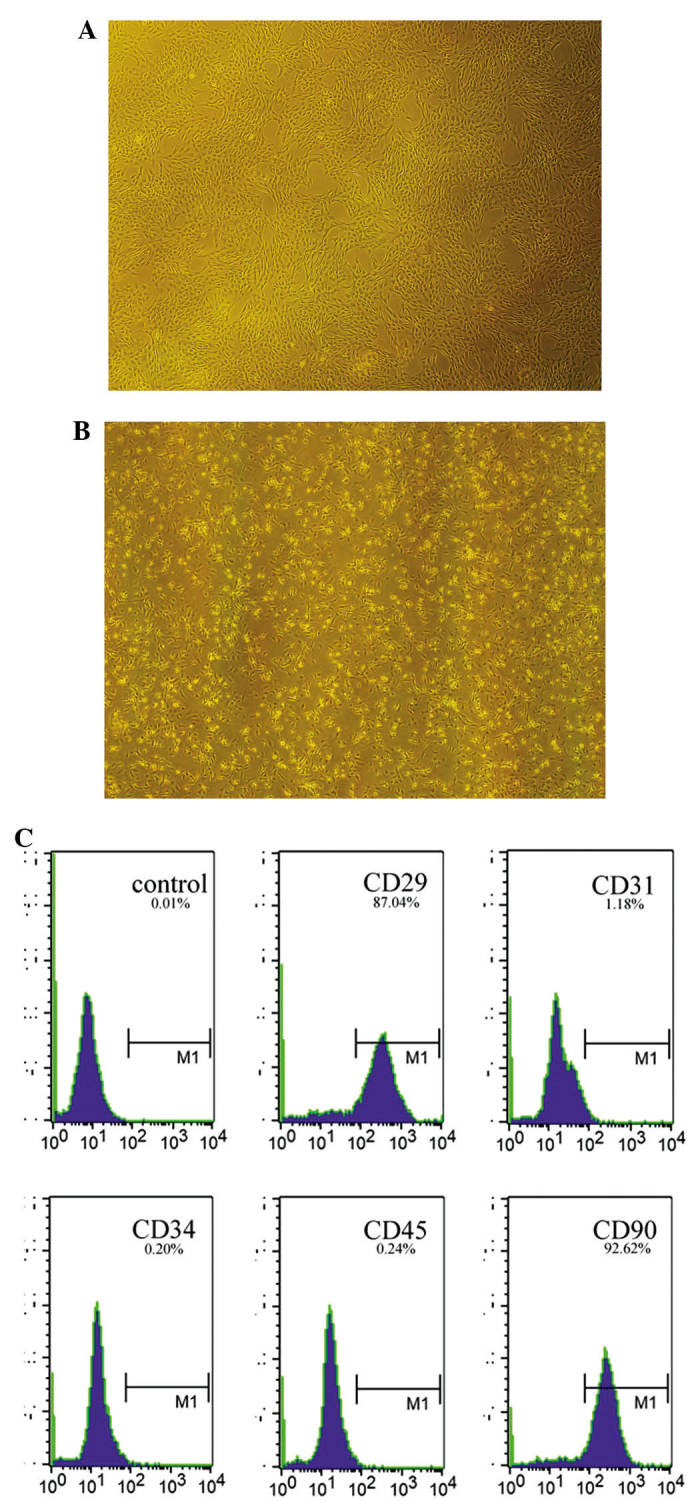
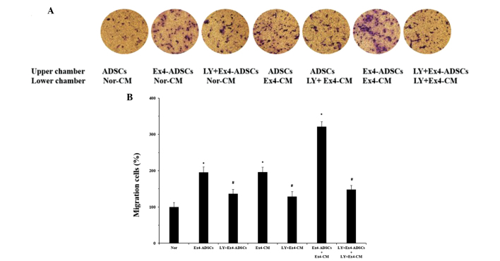
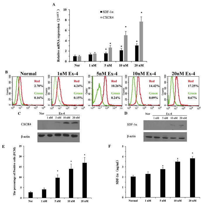
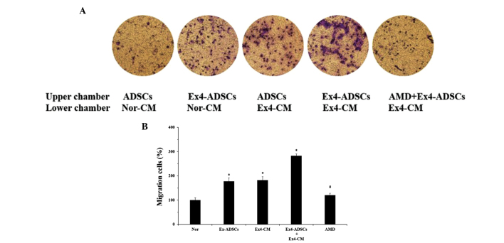
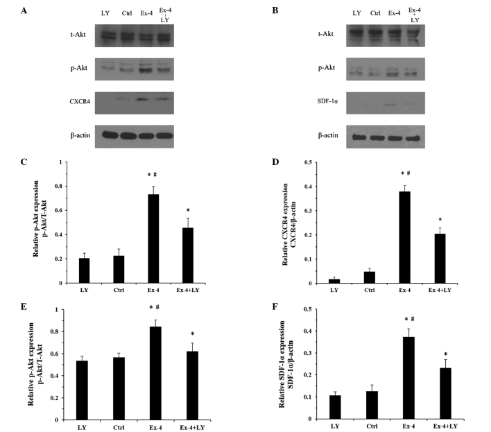
Adipose‑derived stem cells (ADSCs) are considered a suitable source of cells for the repair of tissue following acute myocardial infarction (AMI); however, the transplantation efficiency of ADSCs remains low. Therefore, identification of an efficient method to enhance the migration of engrafted cells to the target site is required. The present study used exendin‑4 (Ex‑4), a glucagon‑like peptide‑1 receptor agonist, to optimize the migratory capacity of ADSCs. The aim was to determine the effect and mechanisms of Ex‑4 on the migration of ADSCs to neonatal rat ventricular cardiomyocyte‑derived conditioned medium (NRVC‑CM). The ADSCs and cardiomyocytes were cultured in vitro. Following incubation of the ADSCs with Ex‑4, cell proliferation was measured using an MTT assay and the expression levels of CXC chemokine receptor 4 (CXCR4) were investigated by reverse transctiption quantitative polymerase chain reaction (RT‑qPCR), western blot analysis and flow cytometry. In addition, the expression levels of stromal cell‑derived factor‑1α (SDF‑1α) were evaluated in the NRVC‑CM treated with Ex‑4 by ELISA, RT‑qPCR and western blot analysis. The migration of the ADSCs to the NRVC‑CM was examined using a Transwell assay. Changes in the protein expression levels of phosphorylated (p‑)Akt were examined in the two types of cell by western blot analysis. The results suggested that Ex‑4 promoted the proliferation and expression of CXCR4 in the ADSCs, increased the secretion of SDF‑1α in the cardiomyocytes and increased the expression levels of p‑Akt in both cells. However, the alterations to the SDF‑1α/CXCR4 cascade in the cells were abrogated following pretreatment with LY‑294002, a phosphoinositide 3‑kinase(PI3K) inhibitor. Furthermore, a Transwell migration assay revealed marked translocation of the ADSCs through the membranes, towards the NRVC‑CM, following treatment with Ex‑4. However, these effects were reduced significantly by pretreatment of the cells with the SDF‑1α/CXCR4 cascade antagonist, AMD3100, and the PI3K inhibitor, LY‑294002. These results indicated that Ex‑4 augmented the SDF‑1α/CXCR4 cascade by activating the PI3K/Akt pathways in the ADSCs and NRVCs. Furthermore, enhancement of the PI3K/Akt-SDF-1α/CXCR4 pathway may be important in the migratory response of ADSCs to NRVC‑CM in vitro.
脂肪来源干细胞(ADSCs)被认为是急性心肌梗死(AMI)后组织修复的合适细胞来源;然而,ADSCs的移植效率仍然很低。因此,需要确定一种有效的方法来增强植入细胞向靶位点的迁移。本研究使用胰高血糖素样肽-1受体激动剂艾塞那肽-4(Ex-4)来优化ADSCs的迁移能力。目的是确定Ex-4对ADSCs向新生大鼠心室心肌细胞条件培养基(NRVC-CM)迁移的影响及机制。将ADSCs和心肌细胞进行体外培养。用Ex-4孵育ADSCs后,采用MTT法检测细胞增殖,并通过逆转录定量聚合酶链反应(RT-qPCR)、蛋白质免疫印迹分析和流式细胞术研究CXC趋化因子受体4(CXCR4)的表达水平。此外,通过酶联免疫吸附测定(ELISA)、RT-qPCR和蛋白质免疫印迹分析评估用Ex-4处理的NRVC-CM中基质细胞衍生因子-1α(SDF-1α)的表达水平。使用Transwell小室测定法检测ADSCs向NRVC-CM的迁移。通过蛋白质免疫印迹分析检测两种细胞中磷酸化(p-)Akt蛋白表达水平的变化。结果表明,Ex-4促进了ADSCs中CXCR4的增殖和表达,增加了心肌细胞中SDF-1α的分泌,并增加了两种细胞中p-Akt的表达水平。然而,在用磷酸肌醇3-激酶(PI3K)抑制剂LY-294002预处理后,细胞中SDF-!α/CXCR4级联反应的改变被消除。此外,Transwell迁移试验显示,用Ex-4处理后,ADSCs明显穿过膜向NRVC-CM迁移。然而,用SDF-1α/CXCR4级联拮抗剂AMD3100和PI3K抑制剂LY-294002对细胞进行预处理后,这些作用显著降低。这些结果表明,Ex-4通过激活ADSCs和NRVCs中的PI3K/Akt信号通路增强了SDF-1α/CXCR4级联反应。此外,增强PI3K/Akt-SDF-1α/CXCR4信号通路可能在体外ADSCs对NRVC-CM的迁移反应中起重要作用。