Jin Jianliang, Lv Xianhui, Chen Lulu, Zhang Wei, Li Jinbo, Wang Qian, Wang Rong, Lu Xiang, Miao Dengshun
The State Key Laboratory of Reproductive Medicine, Department of Anatomy, Histology and Embryology, Nanjing Medical University, Nanjing, China.
Aging Cell. 2014 Oct;13(5):797-809. doi: 10.1111/acel.12236. Epub 2014 Jun 11.
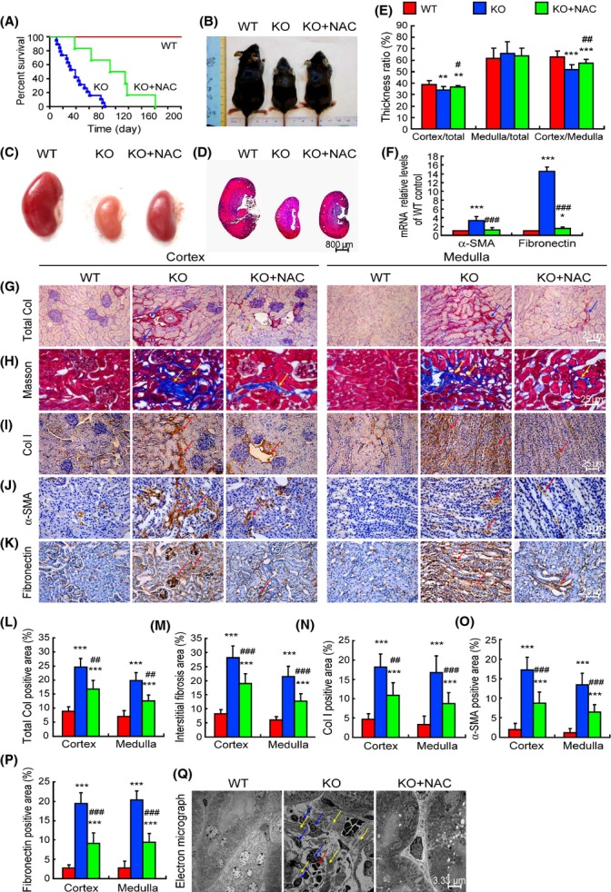
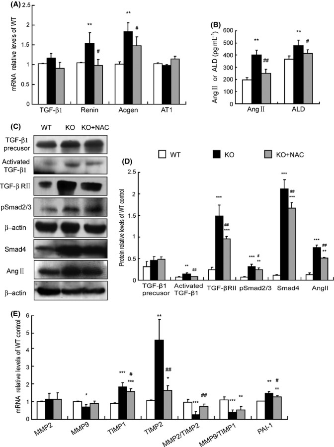
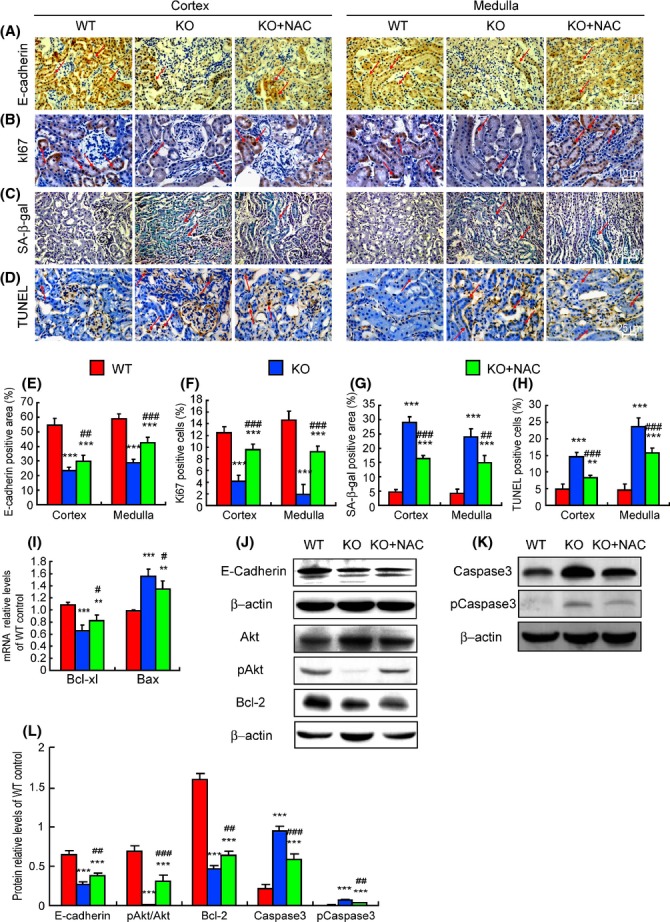
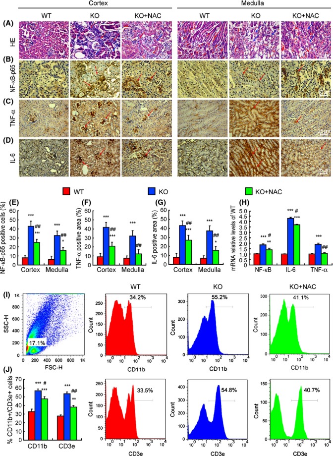
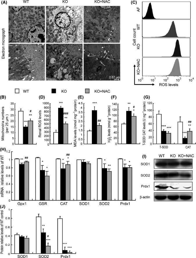
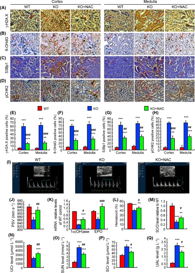
To determine whether Bmi-1 deficiency could lead to renal tubulointerstitial injury by mitochondrial dysfunction and increased oxidative stress in the kidney, 3-week-old Bmi-1(-/-) mice were treated with the antioxidant N-acetylcysteine (NAC, 1 mg mL(-1) ) in their drinking water, or pyrro-quinoline quinone (PQQ, 4 mg kg(-1) diet) in their diet for 2 weeks, and their renal phenotypes were compared with vehicle-treated Bmi1(-/-) and wild-type mice. Bmi-1 was knocked down in human renal proximal tubular epithelial (HK2) cells which were treated with 1 mm NAC for 72 or 96 h, and their phenotypes were compared with control cells. Five-week-old vehicle-treated Bmi-1(-/-) mice displayed renal interstitial fibrosis, tubular atrophy, and severe renal function impairment with decreased renal cell proliferation, increased renal cell apoptosis and senescence, and inflammatory cell infiltration. Impaired mitochondrial structure, decreased mitochondrial numbers, and increased oxidative stress occurred in Bmi-1(-/-) mice; subsequently, this caused DNA damage, the activation of TGF-β1/Smad signaling, and the imbalance between extracellular matrix synthesis and degradation. Oxidative stress-induced epithelial-to-mesenchymal transition of renal tubular epithelial cells was enhanced in Bmi-1 knocked down HK2 cells. All phenotypic alterations caused by Bmi-1 deficiency were ameliorated by antioxidant treatment. These findings indicate that Bmi-1 plays a critical role in protection from renal tubulointerstitial injury by maintaining redox balance and will be a novel therapeutic target for preventing renal tubulointerstitial injury.
为了确定Bmi-1基因缺陷是否会通过线粒体功能障碍和肾脏氧化应激增加导致肾小管间质损伤,给3周龄的Bmi-1(-/-)小鼠饮用含抗氧化剂N-乙酰半胱氨酸(NAC,1 mg mL(-1))的水,或在其饮食中添加吡咯喹啉醌(PQQ,4 mg kg(-1)),持续2周,并将它们的肾脏表型与用赋形剂处理的Bmi1(-/-)小鼠和野生型小鼠进行比较。在人肾近端小管上皮(HK2)细胞中敲低Bmi-1基因,并用1 mM NAC处理72或96小时,然后将其表型与对照细胞进行比较。5周龄用赋形剂处理的Bmi-1(-/-)小鼠表现出肾间质纤维化、肾小管萎缩和严重的肾功能损害,肾细胞增殖减少,肾细胞凋亡和衰老增加,以及炎性细胞浸润。Bmi-1(-/-)小鼠出现线粒体结构受损、线粒体数量减少和氧化应激增加;随后,这导致DNA损伤、TGF-β1/Smad信号通路激活以及细胞外基质合成与降解失衡。在敲低Bmi-1基因的HK2细胞中,氧化应激诱导的肾小管上皮细胞上皮-间质转化增强。抗氧化剂处理改善了Bmi-1基因缺陷引起的所有表型改变。这些发现表明,Bmi-1通过维持氧化还原平衡在保护免受肾小管间质损伤中起关键作用,将成为预防肾小管间质损伤的新治疗靶点。