Sugi Haruo, Chaen Shigeru, Kobayashi Takakazu, Abe Takahiro, Kimura Kazushige, Saeki Yasutake, Ohnuki Yoshiki, Miyakawa Takuya, Tanokura Masaru, Sugiura Seiryo
Department of Physiology, School of Medicine, Teikyo University, Tokyo, Japan.
Department of Integrated Sciences in Physics and Biology, College of Humanities and Sciences, Nihon University, Tokyo, Japan.
PLoS One. 2014 Jun 11;9(2):e93272. doi: 10.1371/journal.pone.0093272. eCollection 2014.
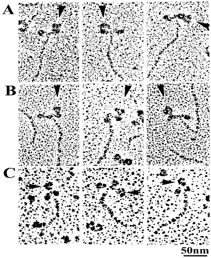
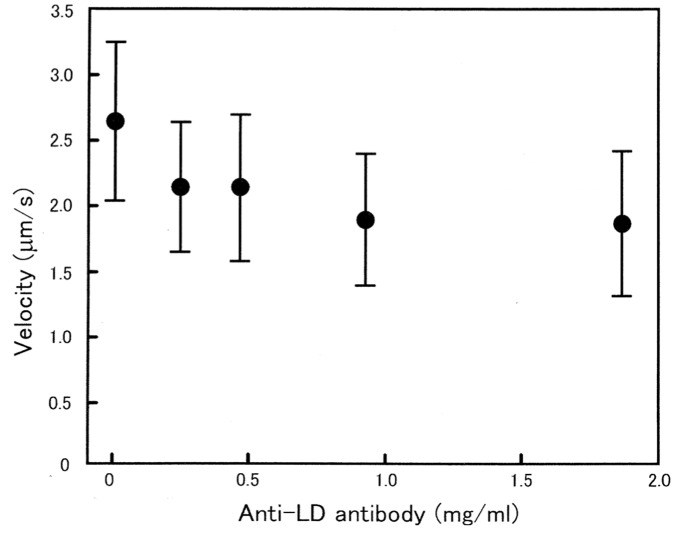
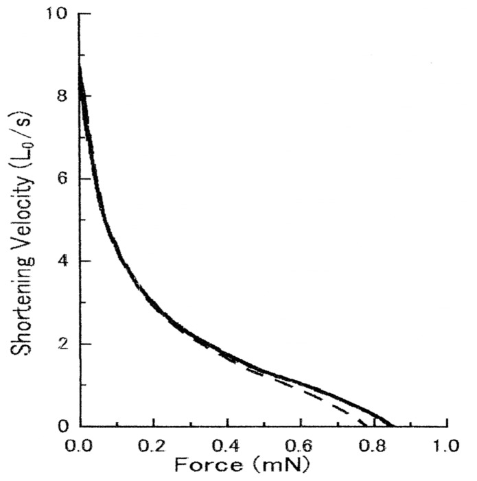
Muscle contraction results from attachment-detachment cycles between myosin heads extending from myosin filaments and actin filaments. It is generally believed that a myosin head first attaches to actin, undergoes conformational changes to produce force and motion in muscle, and then detaches from actin. Despite extensive studies, the molecular mechanism of myosin head conformational changes still remains to be a matter for debate and speculation. The myosin head consists of catalytic (CAD), converter (CVD) and lever arm (LD) domains. To give information about the role of these domains in the myosin head performance, we have examined the effect of three site-directed antibodies to the myosin head on in vitro ATP-dependent actin-myosin sliding and Ca2+-activated contraction of muscle fibers. Antibody 1, attaching to junctional peptide between 50K and 20K heavy chain segments in the CAD, exhibited appreciable effects neither on in vitro actin-myosin sliding nor muscle fiber contraction. Since antibody 1 covers actin-binding sites of the CAD, one interpretation of this result is that rigor actin-myosin linkage is absent or at most a transient intermediate in physiological actin-myosin cycling. Antibody 2, attaching to reactive lysine residue in the CVD, showed a marked inhibitory effect on in vitro actin-myosin sliding without changing actin-activated myosin head (S1) ATPase activity, while it showed no appreciable effect on muscle contraction. Antibody 3, attaching to two peptides of regulatory light chains in the LD, had no significant effect on in vitro actin-myosin sliding, while it reduced force development in muscle fibers without changing MgATPase activity. The above definite differences in the effect of antibodies 2 and 3 between in vitro actin-myosin sliding and muscle contraction can be explained by difference in experimental conditions; in the former, myosin heads are randomly oriented on a glass surface, while in the latter myosin heads are regularly arranged within filament-lattice structures.
肌肉收缩源于从肌球蛋白丝伸出的肌球蛋白头部与肌动蛋白丝之间的附着 - 分离循环。一般认为,肌球蛋白头部首先附着于肌动蛋白,经历构象变化以在肌肉中产生力和运动,然后从肌动蛋白上分离。尽管进行了广泛研究,但肌球蛋白头部构象变化的分子机制仍存在争议和猜测。肌球蛋白头部由催化结构域(CAD)、转换器结构域(CVD)和杠杆臂结构域(LD)组成。为了了解这些结构域在肌球蛋白头部功能中的作用,我们研究了针对肌球蛋白头部的三种位点特异性抗体对体外ATP依赖性肌动蛋白 - 肌球蛋白滑动以及肌肉纤维Ca2 +激活收缩的影响。抗体1附着于CAD中50K和20K重链片段之间的连接肽,对体外肌动蛋白 - 肌球蛋白滑动和肌肉纤维收缩均未表现出明显影响。由于抗体1覆盖了CAD的肌动蛋白结合位点,这一结果的一种解释是,在生理肌动蛋白 - 肌球蛋白循环中不存在强直肌动蛋白 - 肌球蛋白连接,或者至多是一种短暂的中间体。抗体2附着于CVD中的反应性赖氨酸残基,对体外肌动蛋白 - 肌球蛋白滑动表现出显著抑制作用,而不改变肌动蛋白激活的肌球蛋白头部(S1)ATP酶活性,但对肌肉收缩未表现出明显影响。抗体3附着于LD中调节轻链的两个肽段,对体外肌动蛋白 - 肌球蛋白滑动无显著影响,但在不改变MgATP酶活性的情况下降低了肌肉纤维中的力产生。抗体2和3在体外肌动蛋白 - 肌球蛋白滑动和肌肉收缩方面作用的上述明确差异可以通过实验条件的差异来解释;在前者中,肌球蛋白头部随机取向于玻璃表面,而在后者中,肌球蛋白头部规则排列在细丝 - 晶格结构内。