• 文献检索
  • 文档翻译
  • 深度研究
  • 学术资讯
  • Suppr Zotero 插件Zotero 插件
  • 邀请有礼
  • 套餐&价格
  • 历史记录
应用&插件
Suppr Zotero 插件Zotero 插件浏览器插件Mac 客户端Windows 客户端微信小程序
定价
高级版会员购买积分包购买API积分包
服务
文献检索文档翻译深度研究API 文档MCP 服务
关于我们
关于 Suppr公司介绍联系我们用户协议隐私条款
关注我们

Suppr 超能文献

核心技术专利:CN118964589B侵权必究
粤ICP备2023148730 号-1Suppr @ 2026

文献检索

告别复杂PubMed语法,用中文像聊天一样搜索,搜遍4000万医学文献。AI智能推荐,让科研检索更轻松。

立即免费搜索

文件翻译

保留排版,准确专业,支持PDF/Word/PPT等文件格式,支持 12+语言互译。

免费翻译文档

深度研究

AI帮你快速写综述,25分钟生成高质量综述,智能提取关键信息,辅助科研写作。

立即免费体验

年轻患者非典型黑素细胞增生和黑色素瘤的荧光原位杂交分析

Fluorescence in situ hybridization analysis of atypical melanocytic proliferations and melanoma in young patients.

作者信息

DeMarchis Emilia H, Swetter Susan M, Jennings Charay D, Kim Jinah

机构信息

School of Medicine, Stanford University, Stanford, California, USA.

出版信息

Pediatr Dermatol. 2014 Sep-Oct;31(5):561-9. doi: 10.1111/pde.12382. Epub 2014 Jun 13.

DOI:10.1111/pde.12382
PMID:24924836
原文链接:https://pmc.ncbi.nlm.nih.gov/articles/PMC4282368/
Abstract

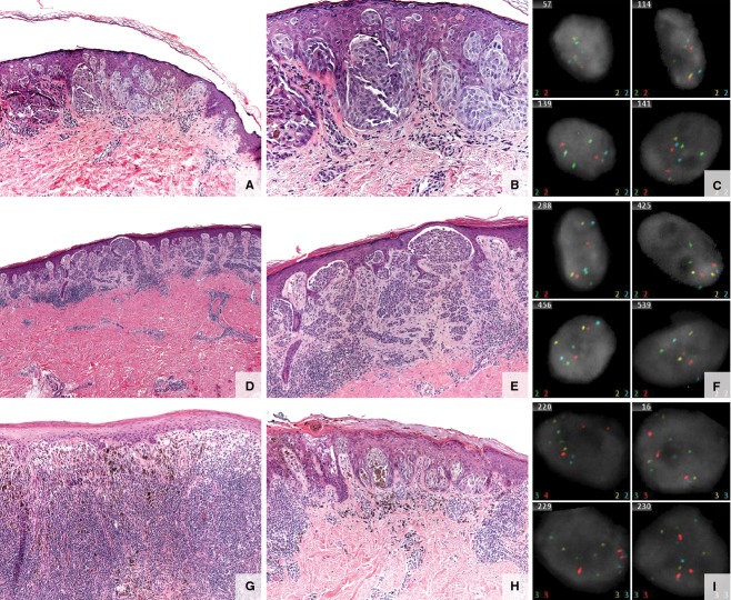
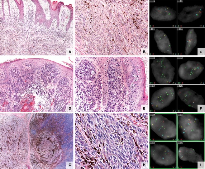
Morphologic heterogeneity among melanocytic proliferations is a common challenge in the diagnosis of melanoma. In particular, atypical melanocytic lesions in children, adolescents, and young adults may be difficult to classify because of significant morphologic overlap with melanoma. Recently a four-probe fluorescence in situ hybridization (FISH) protocol to detect chromosomal abnormalities in chromosomes 6 and 11 has shown promise for improving the classification of melanocytic lesions. We sought to determine the correlation between FISH results, morphology, and clinical outcomes in a series of challenging melanocytic proliferations in young patients. We retrospectively performed the standard four-probe FISH analysis on 21 melanocytic neoplasms from 21 patients younger than 25 years of age (range 5-25 years, mean 14.6 years) from Stanford University Medical Center who were prospectively followed for a median of 51 months (range 1-136 months). The study cohort included patients with 5 confirmed melanomas, 2 melanocytic tumors of uncertain malignant potential (MelTUMPs), 10 morphologically challenging atypical Spitz tumors (ASTs), and 4 typical Spitz nevi. FISH detected chromosomal aberrations in all five melanomas and in one MelTUMP, in which the patient developed subsequent lymph node and distant metastasis. All 10 ASTs, 4 Spitz nevi, and 1 of 2 MelTUMPs were negative for significant gains or losses in chromosomes 6 and 11q. Our findings demonstrated a strong correlation between positive FISH results and the histomorphologic impression of melanoma. This finding was also true for the MelTUMP with poor clinical outcome. Therefore FISH may serve as a helpful adjunct in the classification of controversial melanocytic tumors in young patients.

摘要

黑素细胞增殖的形态学异质性是黑色素瘤诊断中常见的挑战。特别是儿童、青少年和年轻成年人中的非典型黑素细胞病变,由于与黑色素瘤存在显著的形态学重叠,可能难以分类。最近,一种用于检测6号和11号染色体染色体异常的四探针荧光原位杂交(FISH)方案显示出有望改善黑素细胞病变的分类。我们试图确定在一系列年轻患者具有挑战性的黑素细胞增殖中FISH结果、形态学和临床结果之间的相关性。我们对来自斯坦福大学医学中心的21例年龄小于25岁(范围5 - 25岁,平均14.6岁)的患者的21个黑素细胞肿瘤进行了回顾性标准四探针FISH分析,这些患者接受了中位51个月(范围1 - 136个月)的前瞻性随访。研究队列包括5例确诊黑色素瘤患者、2例恶性潜能不确定的黑素细胞肿瘤(MelTUMPs)、10例形态学上具有挑战性的非典型斯皮茨肿瘤(ASTs)和4例典型斯皮茨痣。FISH在所有5例黑色素瘤和1例MelTUMP中检测到染色体畸变,该例患者随后发生了淋巴结和远处转移。所有10例ASTs、4例斯皮茨痣以及2例MelTUMPs中的1例在6号染色体和11q染色体上均未出现显著的增益或缺失。我们的研究结果表明FISH阳性结果与黑色素瘤的组织形态学印象之间存在很强的相关性。这一发现对于临床结果较差的MelTUMP也是如此。因此,FISH可能有助于对年轻患者中有争议的黑素细胞肿瘤进行分类。

https://cdn.ncbi.nlm.nih.gov/pmc/blobs/c192/4282368/2f489549bbb5/pde0031-0561-f2.jpg
https://cdn.ncbi.nlm.nih.gov/pmc/blobs/c192/4282368/c812de5e5989/pde0031-0561-f1.jpg
https://cdn.ncbi.nlm.nih.gov/pmc/blobs/c192/4282368/2f489549bbb5/pde0031-0561-f2.jpg
https://cdn.ncbi.nlm.nih.gov/pmc/blobs/c192/4282368/c812de5e5989/pde0031-0561-f1.jpg
https://cdn.ncbi.nlm.nih.gov/pmc/blobs/c192/4282368/2f489549bbb5/pde0031-0561-f2.jpg

相似文献

1
Fluorescence in situ hybridization analysis of atypical melanocytic proliferations and melanoma in young patients.年轻患者非典型黑素细胞增生和黑色素瘤的荧光原位杂交分析
Pediatr Dermatol. 2014 Sep-Oct;31(5):561-9. doi: 10.1111/pde.12382. Epub 2014 Jun 13.
2
Fluorescence in situ hybridization (FISH) analysis of melanocytic nevi and melanomas: sensitivity, specificity, and lack of association with sentinel node status.黑素细胞痣和黑色素瘤的荧光原位杂交(FISH)分析:敏感性、特异性以及与前哨淋巴结状态无关
Int J Surg Pathol. 2012 Oct;20(5):434-40. doi: 10.1177/1066896912445923. Epub 2012 May 4.
3
Fluorescence in situ hybridization as an ancillary method for the distinction of desmoplastic melanomas from sclerosing melanocytic nevi.荧光原位杂交作为鉴别促纤维增生性黑色素瘤与硬化性黑素细胞痣的辅助方法。
J Cutan Pathol. 2011 Apr;38(4):329-34. doi: 10.1111/j.1600-0560.2010.01666.x.
4
Copy number variations and clinical outcome in atypical spitz tumors.非典型 Spitz 肿瘤中的拷贝数变异与临床结局。
Am J Surg Pathol. 2011 Feb;35(2):243-52. doi: 10.1097/PAS.0b013e31820393ee.
5
Diagnosis of cutaneous melanocytic tumours by four-colour fluorescence in situ hybridisation.通过四色荧光原位杂交诊断皮肤黑素细胞肿瘤
Pathology. 2009;41(4):383-7. doi: 10.1080/00313020902915875.
6
Proliferation indices correlate with diagnosis and metastasis in diagnostically challenging melanocytic tumors.增殖指数与诊断困难的黑素细胞肿瘤的诊断及转移相关。
Hum Pathol. 2016 Jul;53:73-81. doi: 10.1016/j.humpath.2016.02.019. Epub 2016 Mar 19.
7
Update on fluorescence in situ hybridization in melanoma: state of the art.黑色素瘤荧光原位杂交的最新进展:现状。
Arch Pathol Lab Med. 2011 Jul;135(7):830-7. doi: 10.5858/2011-0048-RAIR.1.
8
Melanoma and melanocytic tumors of uncertain malignant potential in children, adolescents and young adults--the Stanford experience 1995-2008.儿童、青少年及青年的黑色素瘤和恶性潜能不确定的黑素细胞肿瘤——斯坦福大学1995 - 2008年的经验
Pediatr Dermatol. 2010 May-Jun;27(3):244-54. doi: 10.1111/j.1525-1470.2009.01078.x. Epub 2010 Apr 9.
9
PRAME expression by immunohistochemistry and reverse transcription quantitative PCR in conjunctival melanocytic lesions-a comprehensive clinicopathologic study of 202 cases and correlation of cytogenetics with PRAME expression in challenging conjunctival melanocytic lesions.通过免疫组织化学和逆转录定量PCR检测PRAME在结膜黑素细胞性病变中的表达——202例综合临床病理研究以及在具有挑战性的结膜黑素细胞性病变中细胞遗传学与PRAME表达的相关性
Hum Pathol. 2023 Apr;134:1-18. doi: 10.1016/j.humpath.2023.02.002. Epub 2023 Feb 16.
10
Deep penetrating nevus-like borderline tumors: A unique subset of ambiguous melanocytic tumors with malignant potential and normal cytogenetics.深部穿透性痣样交界性肿瘤:一类具有恶性潜能且细胞遗传学正常的特殊含混不清黑素细胞肿瘤亚型。
Eur J Dermatol. 2014 Sep-Oct;24(5):594-602. doi: 10.1684/ejd.2014.2393.

引用本文的文献

1
Melanocytic tumors with spitzoid morphology: correlation of clinicopathologic features and FISH analysis.梭形细胞/具有斯皮茨样形态特征的黑色素细胞肿瘤:临床病理特征与 FISH 分析的相关性。
Turk J Med Sci. 2023 Aug;53(4):924-933. doi: 10.55730/1300-0144.5656. Epub 2023 Aug 18.
2
Clinical features and outcomes of spitzoid proliferations in children and adolescents.儿童和青少年 Spitz 样增生的临床特征和结局。
Br J Dermatol. 2019 Aug;181(2):366-372. doi: 10.1111/bjd.17450. Epub 2019 Feb 10.
3
Spitzoid melanoma of childhood: a case series and review.

本文引用的文献

1
Comparative analysis of atypical spitz tumors with heterozygous versus homozygous 9p21 deletions for clinical outcomes, histomorphology, BRAF mutation, and p16 expression.杂合性与纯合性 9p21 缺失的非典型 Spitz 肿瘤的临床结局、组织形态学、BRAF 突变和 p16 表达的对比分析。
Am J Surg Pathol. 2014 May;38(5):638-45. doi: 10.1097/PAS.0000000000000160.
2
Pediatric melanoma: analysis of an international registry.小儿黑色素瘤:国际登记处分析。
Cancer. 2013 Nov 15;119(22):4012-9. doi: 10.1002/cncr.28289. Epub 2013 Sep 10.
3
Controversies in the evaluation and management of atypical melanocytic proliferations in children, adolescents, and young adults.
儿童斯皮茨样黑色素瘤:病例系列报道及文献复习
Melanoma Manag. 2015 May;2(2):121-125. doi: 10.2217/mmt.15.6. Epub 2015 May 18.
4
Atypical Melanocytic Proliferations: A Review of the Literature.非典型黑素细胞增生:文献综述
Dermatol Surg. 2018 Feb;44(2):159-174. doi: 10.1097/DSS.0000000000001367.
儿童、青少年和青年人群中非典型黑素细胞增生的评估和处理中的争议。
J Natl Compr Canc Netw. 2013 Jun 1;11(6):679-86. doi: 10.6004/jnccn.2013.0087.
4
Risk assessment for atypical spitzoid melanocytic neoplasms using FISH to identify chromosomal copy number aberrations.使用 FISH 技术识别染色体拷贝数异常进行非典型 Spitz 样黑素细胞肿瘤的风险评估。
Am J Surg Pathol. 2013 May;37(5):676-84. doi: 10.1097/PAS.0b013e3182753de6.
5
Update in molecular diagnostics in melanocytic neoplasms.黑素细胞肿瘤的分子诊断学进展。
Adv Anat Pathol. 2012 Nov;19(6):410-6. doi: 10.1097/PAP.0b013e318271a5cb.
6
Fluorescence in situ hybridization for ambiguous melanocytic tumors.原位杂交荧光检测用于诊断不明确的黑色素细胞肿瘤。
Histol Histopathol. 2012 Dec;27(12):1539-42. doi: 10.14670/HH-27.1539.
7
Differentiation of malignant melanoma from benign nevus using a novel genomic microarray with low specimen requirements.使用新型基因组微阵列,通过低标本要求区分恶性黑色素瘤和良性痣。
Arch Pathol Lab Med. 2012 Aug;136(8):947-55. doi: 10.5858/arpa.2011-0330-OA.
8
A highly specific and discriminatory FISH assay for distinguishing between benign and malignant melanocytic neoplasms.一种高度特异和有鉴别能力的 FISH 检测方法,用于区分良性和恶性黑素细胞肿瘤。
Am J Surg Pathol. 2012 Jun;36(6):808-17. doi: 10.1097/PAS.0b013e31824b1efd.
9
Management of melanomas in children and young adults.儿童和青年黑色素瘤的管理
J Pediatr Hematol Oncol. 2012 May;34 Suppl 2:S51-4. doi: 10.1097/MPH.0b013e31824e3852.
10
Correlation between histologic assessment and fluorescence in situ hybridization using MelanoSITE in evaluation of histologically ambiguous melanocytic lesions.利用 MelanoSITE 在组织学上不确定的黑色素细胞病变中的组织学评估与荧光原位杂交的相关性。
Arch Pathol Lab Med. 2012 Dec;136(12):1571-9. doi: 10.5858/arpa.2011-0673-OA. Epub 2012 Apr 5.