• 文献检索
  • 文档翻译
  • 深度研究
  • 学术资讯
  • Suppr Zotero 插件Zotero 插件
  • 邀请有礼
  • 套餐&价格
  • 历史记录
应用&插件
Suppr Zotero 插件Zotero 插件浏览器插件Mac 客户端Windows 客户端微信小程序
定价
高级版会员购买积分包购买API积分包
服务
文献检索文档翻译深度研究API 文档MCP 服务
关于我们
关于 Suppr公司介绍联系我们用户协议隐私条款
关注我们

Suppr 超能文献

核心技术专利:CN118964589B侵权必究
粤ICP备2023148730 号-1Suppr @ 2026

文献检索

告别复杂PubMed语法,用中文像聊天一样搜索,搜遍4000万医学文献。AI智能推荐,让科研检索更轻松。

立即免费搜索

文件翻译

保留排版,准确专业,支持PDF/Word/PPT等文件格式,支持 12+语言互译。

免费翻译文档

深度研究

AI帮你快速写综述,25分钟生成高质量综述,智能提取关键信息,辅助科研写作。

立即免费体验

心外膜为心脏再生指明了道路。

The epicardium signals the way towards heart regeneration.

作者信息

Masters Megan, Riley Paul R

机构信息

Department of Physiology Anatomy and Genetics, Sherrington Building, University of Oxford, Oxford OX1 3PT, UK.

出版信息

Stem Cell Res. 2014 Nov;13(3 Pt B):683-92. doi: 10.1016/j.scr.2014.04.007. Epub 2014 Apr 29.

DOI:10.1016/j.scr.2014.04.007
PMID:24933704
原文链接:https://pmc.ncbi.nlm.nih.gov/articles/PMC4241487/
Abstract

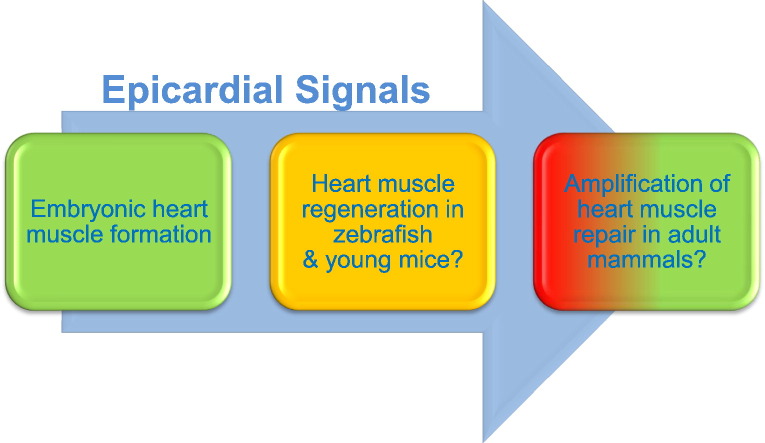
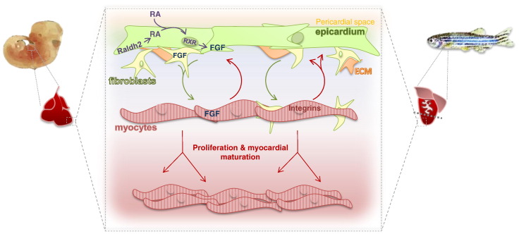
From historical studies of developing chick hearts to recent advances in regenerative injury models, the epicardium has arisen as a key player in heart genesis and repair. The epicardium provides paracrine signals to nurture growth of the developing heart from mid-gestation, and epicardium-derived cells act as progenitors of numerous cardiac cell types. Interference with either process is terminal for heart development and embryogenesis. In adulthood, the dormant epicardium reinstates an embryonic gene programme in response to injury. Furthermore, injury-induced epicardial signalling is essential for heart regeneration in zebrafish. Given these critical roles in development, injury response and heart regeneration, the application of epicardial signals following adult heart injury could offer therapeutic strategies for the treatment of ischaemic heart disease and heart failure.

摘要

从对发育中的鸡心脏的历史研究到再生损伤模型的最新进展,心外膜已成为心脏发生和修复的关键参与者。心外膜提供旁分泌信号,从中孕期开始促进发育中心脏的生长,心外膜衍生细胞作为多种心脏细胞类型的祖细胞。对这两个过程中任何一个的干扰都会导致心脏发育和胚胎发生的终止。在成年期,休眠的心外膜会在损伤时恢复胚胎基因程序。此外,损伤诱导的心外膜信号对于斑马鱼的心脏再生至关重要。鉴于其在发育、损伤反应和心脏再生中的这些关键作用,成年心脏损伤后应用心外膜信号可为缺血性心脏病和心力衰竭的治疗提供治疗策略。

https://cdn.ncbi.nlm.nih.gov/pmc/blobs/17c0/4241487/62296ae6c8be/gr1.jpg
https://cdn.ncbi.nlm.nih.gov/pmc/blobs/17c0/4241487/f3ffec57e947/fx1.jpg
https://cdn.ncbi.nlm.nih.gov/pmc/blobs/17c0/4241487/62296ae6c8be/gr1.jpg
https://cdn.ncbi.nlm.nih.gov/pmc/blobs/17c0/4241487/f3ffec57e947/fx1.jpg
https://cdn.ncbi.nlm.nih.gov/pmc/blobs/17c0/4241487/62296ae6c8be/gr1.jpg

相似文献

1
The epicardium signals the way towards heart regeneration.心外膜为心脏再生指明了道路。
Stem Cell Res. 2014 Nov;13(3 Pt B):683-92. doi: 10.1016/j.scr.2014.04.007. Epub 2014 Apr 29.
2
Regenerative potential of epicardium-derived extracellular vesicles mediated by conserved miRNA transfer.心肌外膜细胞外囊泡通过保守的 miRNA 转移介导的再生潜力。
Cardiovasc Res. 2022 Jan 29;118(2):597-611. doi: 10.1093/cvr/cvab054.
3
The Role of the Epicardium During Heart Development and Repair.心外膜在心脏发育和修复中的作用。
Circ Res. 2020 Jan 31;126(3):377-394. doi: 10.1161/CIRCRESAHA.119.315857. Epub 2020 Jan 30.
4
The epicardium as a hub for heart regeneration.心脏外膜作为心脏再生的中心。
Nat Rev Cardiol. 2018 Oct;15(10):631-647. doi: 10.1038/s41569-018-0046-4.
5
The epicardium in cardiac repair: from the stem cell view.心脏修复中的心外膜:从干细胞的角度看。
Pharmacol Ther. 2011 Jan;129(1):82-96. doi: 10.1016/j.pharmthera.2010.09.002. Epub 2010 Oct 19.
6
Epicardial regeneration is guided by cardiac outflow tract and Hedgehog signalling.心外膜再生受心脏流出道和刺猬信号通路的引导。
Nature. 2015 Jun 11;522(7555):226-230. doi: 10.1038/nature14325. Epub 2015 May 4.
7
Cardiac regeneration: epicardial mediated repair.心脏再生:心外膜介导的修复
Proc Biol Sci. 2015 Dec 22;282(1821):20152147. doi: 10.1098/rspb.2015.2147.
8
Epicardial HDAC3 Promotes Myocardial Growth Through a Novel MicroRNA Pathway.心外膜 HDAC3 通过新型 microRNA 通路促进心肌生长。
Circ Res. 2022 Jul 8;131(2):151-164. doi: 10.1161/CIRCRESAHA.122.320785. Epub 2022 Jun 20.
9
The ontogeny, activation and function of the epicardium during heart development and regeneration.心外膜在心脏发育和再生过程中的个体发生、激活和功能。
Development. 2018 Mar 28;145(7):dev155994. doi: 10.1242/dev.155994.
10
Epicardium and myocardium separate from a common precursor pool by crosstalk between bone morphogenetic protein- and fibroblast growth factor-signaling pathways.心外膜和心肌通过骨形态发生蛋白信号通路与成纤维细胞生长因子信号通路之间的相互作用,从共同的前体细胞库中分离出来。
Circ Res. 2009 Aug 28;105(5):431-41. doi: 10.1161/CIRCRESAHA.109.203083. Epub 2009 Jul 23.

引用本文的文献

1
Cardiac Regeneration and Repair in Zebrafish and Mammalian Models.斑马鱼和哺乳动物模型中的心脏再生与修复
Curr Cardiol Rep. 2025 Jun 17;27(1):95. doi: 10.1007/s11886-025-02235-6.
2
BubR1 Controls Heart Development by Promoting Expression of Cardiogenesis Regulators.BubR1通过促进心脏发生调节因子的表达来控制心脏发育。
J Am Heart Assoc. 2025 Mar 18;14(6):e038286. doi: 10.1161/JAHA.124.038286. Epub 2025 Mar 7.
3
CD63 as novel target for nanoemulsion-based F MRI imaging and drug delivery to activated cardiac fibroblasts.CD63作为基于纳米乳剂的功能磁共振成像以及向活化心肌成纤维细胞递送药物的新靶点。

本文引用的文献

1
Harnessing the power of dividing cardiomyocytes.利用分裂心肌细胞的力量。
Glob Cardiol Sci Pract. 2013 Nov 1;2013(3):212-21. doi: 10.5339/gcsp.2013.29. eCollection 2013.
2
Differential expression of embryonic epicardial progenitor markers and localization of cardiac fibrosis in adult ischemic injury and hypertensive heart disease.胚胎心外膜祖细胞标志物的差异表达及成年缺血性损伤和高血压性心脏病中心脏纤维化的定位。
J Mol Cell Cardiol. 2013 Dec;65:108-19. doi: 10.1016/j.yjmcc.2013.10.005. Epub 2013 Oct 17.
3
Depletion of retinoic acid receptors initiates a novel positive feedback mechanism that promotes teratogenic increases in retinoic acid.
Theranostics. 2025 Jan 1;15(1):1-18. doi: 10.7150/thno.96990. eCollection 2025.
4
LncRNAs in the Domain Are Essential for Mid-Embryonic Heart Development.领域中的 lncRNAs 对胚胎中期心脏发育至关重要。
Int J Mol Sci. 2024 Jul 26;25(15):8184. doi: 10.3390/ijms25158184.
5
Epicardial EMT and cardiac repair: an update.心外膜 EMT 与心脏修复:研究进展。
Stem Cell Res Ther. 2024 Jul 19;15(1):219. doi: 10.1186/s13287-024-03823-z.
6
Distinct features of the regenerating heart uncovered through comparative single-cell profiling.通过比较单细胞分析揭示再生心脏的独特特征。
Biol Open. 2024 Apr 15;13(4). doi: 10.1242/bio.060156. Epub 2024 Apr 5.
7
The Neural Progenitor Cell-Associated Transcription Factor FoxG1 Regulates Cardiac Epicardial Cell Proliferation.神经祖细胞相关转录因子FoxG1调节心脏心外膜细胞增殖。
Stem Cells Int. 2024 Jan 11;2024:8601360. doi: 10.1155/2024/8601360. eCollection 2024.
8
Understanding Epicardial Cell Heterogeneity during Cardiogenesis and Heart Regeneration.了解心脏发生和心脏再生过程中的心外膜细胞异质性。
J Cardiovasc Dev Dis. 2023 Sep 1;10(9):376. doi: 10.3390/jcdd10090376.
9
Distinct features of the regenerating heart uncovered through comparative single-cell profiling.通过比较单细胞分析揭示的再生心脏的独特特征。
bioRxiv. 2023 Jul 12:2023.07.04.547574. doi: 10.1101/2023.07.04.547574.
10
Am80, a retinoic acid receptor agonist, activates the cardiomyocyte cell cycle and enhances engraftment in the heart.Am80,一种维 A 酸受体激动剂,能激活心肌细胞周期,增强心脏中的植入效果。
Stem Cell Reports. 2023 Aug 8;18(8):1672-1685. doi: 10.1016/j.stemcr.2023.06.006. Epub 2023 Jul 13.
视黄酸受体耗竭会引发一种新的正反馈机制,促进视黄酸致畸作用的增加。
PLoS Genet. 2013;9(8):e1003689. doi: 10.1371/journal.pgen.1003689. Epub 2013 Aug 8.
4
Fibronectin is deposited by injury-activated epicardial cells and is necessary for zebrafish heart regeneration.纤维连接蛋白由损伤激活的心脏外膜细胞分泌,并对斑马鱼心脏再生是必需的。
Dev Biol. 2013 Oct 15;382(2):427-35. doi: 10.1016/j.ydbio.2013.08.012. Epub 2013 Aug 26.
5
Adult c-kit(pos) cardiac stem cells are necessary and sufficient for functional cardiac regeneration and repair.成体 c-kit(阳性)心脏干细胞对于功能性心脏再生和修复是必需且充分的。
Cell. 2013 Aug 15;154(4):827-42. doi: 10.1016/j.cell.2013.07.039.
6
A dynamic spatiotemporal extracellular matrix facilitates epicardial-mediated vertebrate heart regeneration.动态时空细胞外基质促进心外膜介导的脊椎动物心脏再生。
Dev Biol. 2013 Oct 15;382(2):457-69. doi: 10.1016/j.ydbio.2013.08.002. Epub 2013 Aug 11.
7
Hippo pathway effector Yap promotes cardiac regeneration.Hippo 通路效应物 yap 促进心脏再生。
Proc Natl Acad Sci U S A. 2013 Aug 20;110(34):13839-44. doi: 10.1073/pnas.1313192110. Epub 2013 Aug 5.
8
Mending broken hearts: cardiac development as a basis for adult heart regeneration and repair.修复破损的心脏:心脏发育为成年心脏再生和修复的基础。
Nat Rev Mol Cell Biol. 2013 Aug;14(8):529-41. doi: 10.1038/nrm3619. Epub 2013 Jul 10.
9
Subepicardial endothelial cells invade the embryonic ventricle wall to form coronary arteries.心外膜内皮细胞侵袭胚胎心室壁形成冠状动脉。
Cell Res. 2013 Sep;23(9):1075-90. doi: 10.1038/cr.2013.83. Epub 2013 Jun 25.
10
Meis1 regulates postnatal cardiomyocyte cell cycle arrest.Meis1 调控心肌细胞出生后的细胞周期停滞。
Nature. 2013 May 9;497(7448):249-253. doi: 10.1038/nature12054. Epub 2013 Apr 17.