Tajima Hidehiro, Kitagawa Hirohisa, Shoji Masatoshi, Watanabe Toshifumi, Nakanuma Shinichi, Okamoto Koichi, Sakai Seisho, Kinoshita Jun, Makino Isamu, Furukawa Hiroyuki, Nakamura Keishi, Hayashi Hironori, Oyama Katsunobu, Inokuchi Masafumi, Nakagawara Hisatoshi, Miyashita Tomoharu, Itoh Hiroshi, Takamura Hiroyuki, Ninomiya Itasu, Fushida Sachio, Fujimura Takashi, Ohta Tetsuo, Satoh Hirohide, Ikeda Hiroko, Harada Kenichi, Nakanuma Yasuni
Department of Gastroenterologic Surgery, Division of Cancer Medicine, Graduate School of Medicine Science, Kanazawa University, Kanazawa, Ishikawa 920-8641, Japan.
Division of Pathology, Kanazawa University Hospital, Kanazawa University, Kanazawa, Ishikawa 920-8641, Japan.
Oncol Lett. 2014 Apr;7(4):1049-1052. doi: 10.3892/ol.2014.1873. Epub 2014 Feb 12.
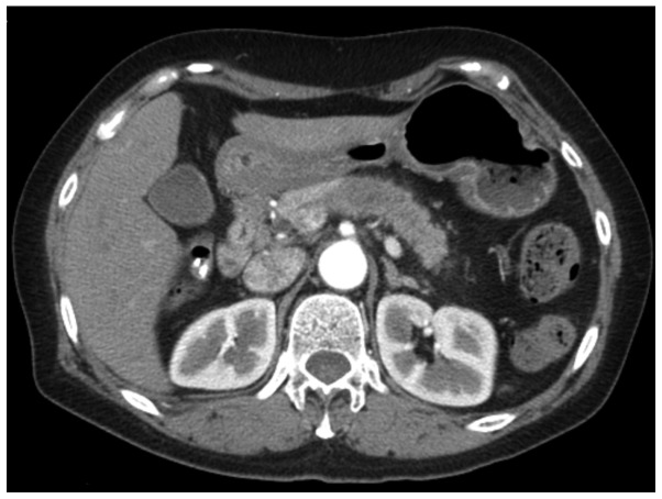
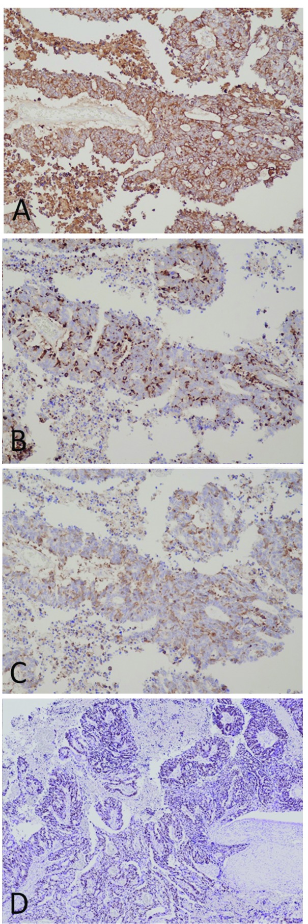
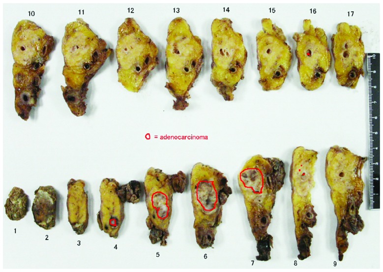
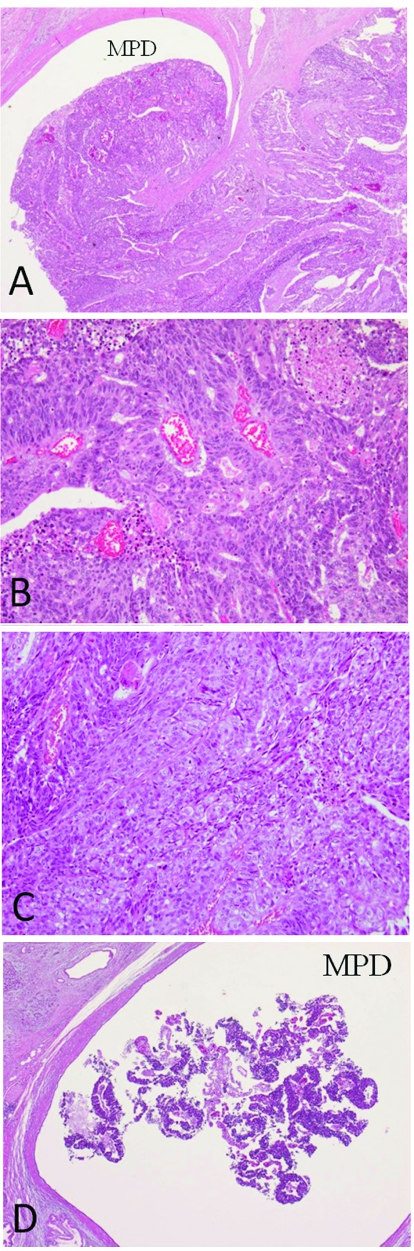
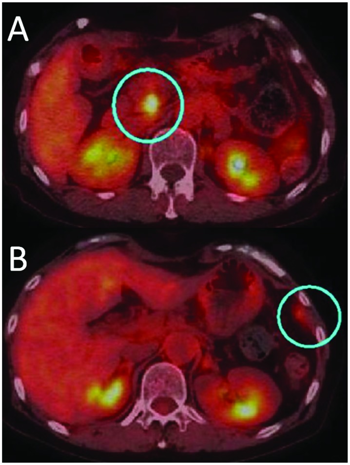
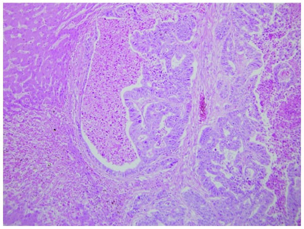
A 61-year-old female with pancreatic body cancer underwent a distal pancreatectomy. The tumor was a moderately- to poorlydifferentiated adenocarcinoma. Tumor growth filled the dilated main pancreatic duct (MPD) and infiltrated the surrounding area. Six months later, metastases to the left diaphragm and MPD of the remnant pancreatic head were detected. Chemoradiotherapy was administered, but the patient succumbed 22 months after surgery. An autopsy demonstrated that a moderately- to poorly-differentiated adenocarcinoma had arisen from the pancreatic head and infiltrated the duodenum and bile duct. Huge liver metastases and multiple peritoneal disseminations were also present. Microscopically, a portion of the tumor had a pseudo-rosette appearance in the adenocarcinoma component, while another section showed characteristics of a neuroendocrine tumor (NET) immunohistochemically. The original surgically-resected tumor also showed NET characteristics immunohistochemically. It is therefore necessary to search for NET components in pancreatic cancer with atypical growth and metastases, even when adenocarcinoma has been diagnosed histologically.
一名61岁的胰腺体部癌女性患者接受了胰腺远端切除术。肿瘤为中分化至低分化腺癌。肿瘤生长充满扩张的主胰管(MPD)并浸润周围区域。六个月后,检测到左膈和残余胰头的MPD转移。给予了放化疗,但患者在术后22个月死亡。尸检显示,中分化至低分化腺癌起源于胰头并浸润十二指肠和胆管。还存在巨大的肝转移和多处腹膜播散。显微镜下,肿瘤的一部分在腺癌成分中呈假菊形团外观,而另一部分免疫组化显示具有神经内分泌肿瘤(NET)的特征。最初手术切除的肿瘤免疫组化也显示出NET特征。因此,即使在组织学上已诊断为腺癌,对于具有非典型生长和转移的胰腺癌,也有必要寻找NET成分。