Sayed Leela, Sangal Sam, Finch Guy
University of Leicester Medical School, Leicester.
Leicester General Hospital, Leicester.
J Surg Case Rep. 2010 Jul 1;2010(5):5. doi: 10.1093/jscr/2010.5.5.
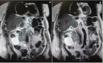
Spontaneous cholecystocutaneous fistula, one of the rarest complications of acute cholecystitis, has been reported in fewer than 25 cases over the past 50 years. Not only is this case rare but interestingly the patient experienced no pain or symptoms consistent with gallbladder pathology leading up to her hospitalisation. Furthermore, laboratory studies, microbiology and computed tomography scanning did not establish a diagnosis until the fistula passed calculi. An 85-year-old lady with multiple co-morbidities presented to the Emergency Department with an erythematous soft and non-tender mass in her right flank. The mass had spontaneously ruptured and was discharging a serous-like material. Prior to further investigation a working diagnosis of an eroding/fungating caecal tumour was made. The lesion continued to discharge over a 3 month period which heralded the passage of 11 small, brown calculi thought to be gallstones. At this point spontaneous cholecystocutaneous fistula was diagnosed and was later confirmed by magnetic resonance imaging cholangiopancreatography.
自发性胆囊皮肤瘘是急性胆囊炎最罕见的并发症之一,在过去50年中报告的病例少于25例。该病例不仅罕见,而且有趣的是,患者在入院前没有出现与胆囊病变相关的疼痛或症状。此外,在瘘管排出结石之前,实验室检查、微生物学检查和计算机断层扫描均未确诊。一名患有多种合并症的85岁女性因右下腹出现一个柔软、无压痛的红斑性肿块而就诊于急诊科。该肿块已自发破裂,排出浆液样物质。在进一步检查之前,初步诊断为盲肠侵蚀性/溃疡性肿瘤。病变在3个月内持续排出物质,随后排出了11颗被认为是胆结石的小褐色结石。此时诊断为自发性胆囊皮肤瘘,后来经磁共振胰胆管造影证实。