Department of Gastroenterology, Post Graduate Institute of Medical Education and Research (PGIMER), Sector 12, Chandigarh 160012, India.
Department of Cytology, Post Graduate Institute of Medical Education and Research (PGIMER), Sector 12, Chandigarh 160012, India.
Endosc Ultrasound. 2013 Apr;2(2):92-5. doi: 10.4103/2303-9027.117693.
Dysphagia as a presenting manifestation of tuberculosis is rare and there is paucity of data on the clinical, endoscopic and endosonographic features of these patients. We present our data related to the features over last four years.
We analyzed retrospectively the clinical, endoscopic, radiological, endosonographic and cytological findings in 14 patients (male: 10; mean age: 37.7 ± 10.4 years) with dysphagia due to tuberculosis presenting to us over last 4 years.
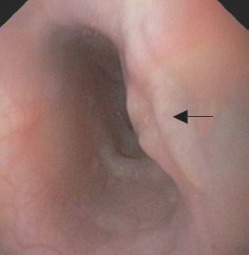
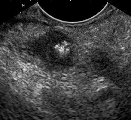
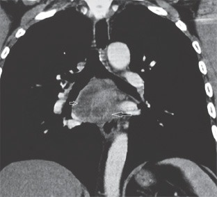
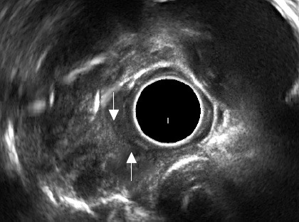
Nine patients (64.3%) had Grade 1 dysphagia, 4 (28.6%) patients had Grade 2 and 1 patient (7.1%) had Grade 3. Mid esophagus was the commonest site of involvement. Endoscopic findings were extrinsic bulge (50%), linear ulcers (28.6%) and pol-ypoidal ulcerated lesion (7.1%). Endoscopic biopsies were inconclusive. Endoscopic ultrasound (EUS) demonstrated mediastinal lymph nodes being responsible for endoscopic bulge and their infiltration into esophageal wall leading on to ulcers. EUS-guided fine needle aspiration from these nodes established diagnosis in all patients.
Dysphagia in tuberculosis is most commonly caused by compression by the surrounding mediastinal lymph nodes. EUS is a useful investigation for assessment of these patients.
以吞咽困难为表现的肺结核较为罕见,有关这些患者的临床、内镜和内镜超声特征的数据也很少。我们报告了过去四年中与这些特征相关的数据。
我们回顾性分析了 14 例(男 10 例;平均年龄 37.7 ± 10.4 岁)因肺结核导致吞咽困难来我院就诊的患者的临床、内镜、影像学、内镜超声和细胞学检查结果。
9 例(64.3%)患者吞咽困难程度为 1 级,4 例(28.6%)患者为 2 级,1 例(7.1%)患者为 3 级。食管中段是最常见的受累部位。内镜下表现为外压性隆起(50%)、线性溃疡(28.6%)和息肉样溃疡性病变(7.1%)。内镜活检结果不明确。内镜超声(EUS)显示纵隔淋巴结是导致内镜下隆起的原因,其浸润食管壁导致溃疡。这些淋巴结的 EUS 引导下细针抽吸活检在所有患者中均确立了诊断。
肺结核引起的吞咽困难最常见的原因是周围纵隔淋巴结的压迫。EUS 是评估这些患者的有用检查方法。