Che Yi-Qun, Wang Shuang, Luo Yang, Wang Jing-Bo, Wang Lu-Hua
Clinical Laboratory, Cancer Institute and Hospital, Chinese Academy of Medical Science and Peking Union Medical College, Beijing 100021, P.R. China.
Department of Radiology, Cancer Institute and Hospital, Chinese Academy of Medical Science and Peking Union Medical College, Beijing 100021, P.R. China.
Oncol Lett. 2014 Jul;8(1):105-110. doi: 10.3892/ol.2014.2064. Epub 2014 Apr 15.
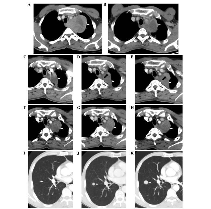
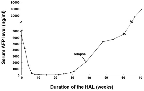
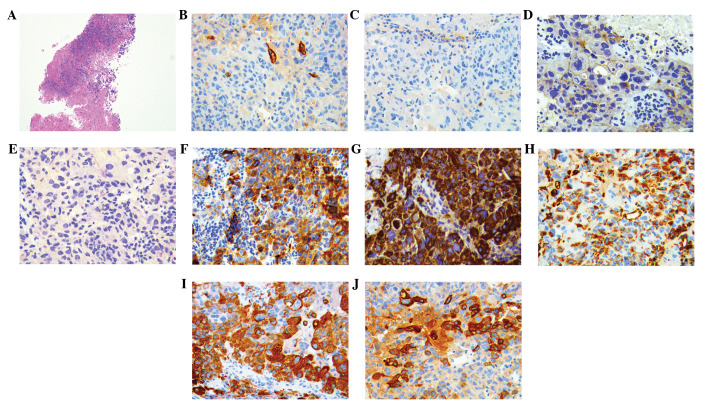
Hepatoid adenocarcinoma of the lung (HAL) is a rare type of lung cancer. Its diagnosis and treatment may be difficult due to the varied presentation; however, immunohistochemical analysis facilitates the diagnosis. The present study presents a case of HAL. The patient was a 48-year-old male who presented with a primary complaint of back pain. A chest computed-tomography scan revealed a lobulated soft-tissue mass that extended from the left lung apex to the middle and posterior mediastinum. The area of the largest cross-section was 7.9×10.0 cm and the lymph nodes did not demonstrate metastasis. Immunohistochemical staining of a transbronchial lung biopsy revealed that the tumor cells were α-fetoprotein (AFP)(positive) and hepatocytes(positive) and a diagnosis of hepatoid carcinoma of the left lung was established. The level of serum AFP, a tumor marker, was elevated (6,283 ng/ml). The patient presented with mediastinal metastases and was classified as stage IIIA (N2); following diagnosis, the patient received concurrent chemoradiation. Subsequent to chemoradiation, the left lung lump with the largest cross-section was 3.3×4.2 cm and the serum AFP had fallen to its lowest level (23.11 ng/ml). However, when the patient relapsed, the serum AFP level elevated markedly (57,800 ng/ml). Furthermore, the nodules of metastasis increased in number and enlarged, with the largest measuring 2.1 cm. The patient succumbed as a result of a lung infection.
肺肝样腺癌(HAL)是一种罕见的肺癌类型。由于其表现多样,其诊断和治疗可能具有挑战性;然而,免疫组化分析有助于诊断。本研究报告了一例HAL病例。患者为48岁男性,主要症状为背痛。胸部计算机断层扫描显示一个分叶状软组织肿块,从左肺尖延伸至纵隔中部和后部。最大横截面面积为7.9×10.0 cm,淋巴结未显示转移。经支气管肺活检的免疫组化染色显示肿瘤细胞α-甲胎蛋白(AFP)阳性且肝细胞阳性,确诊为左肺肝样癌。肿瘤标志物血清AFP水平升高(6283 ng/ml)。患者出现纵隔转移,被分类为IIIA期(N2);诊断后,患者接受同步放化疗。放化疗后,最大横截面的左肺肿块为3.3×4.2 cm,血清AFP降至最低水平(23.11 ng/ml)。然而,当患者复发时,血清AFP水平显著升高(57800 ng/ml)。此外,转移结节数量增加且增大,最大者直径为2.1 cm。患者因肺部感染死亡。