Singh Koijam Sashikumar, Jayachandran S
Department of Oral Medicine and Radiology, Dental College, RIMS, Imphal, Manipur, Tamil Nadu, India.
Department of Oral Medicine and Radiology, Tamil Nadu Government Dental College and Hospital, Chennai, Tamil Nadu, India.
Contemp Clin Dent. 2014 Apr;5(2):166-9. doi: 10.4103/0976-237X.132306.
The objective of the following study is to evaluate the usefulness of ultrasonography (USG) in comparison with conventional radiography and computed tomography (CT) scan in the diagnosis of zygomatic arch and mandibular fractures.
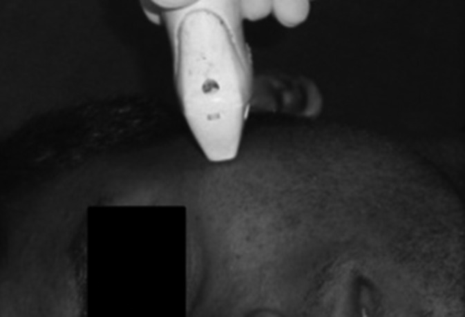
A total of 40 patients with suspected fracture of the zygomatic arch and/or mandibular fractures were included in the study. Two groups (one for zygomatic arch fractures and the other for mandibular fractures) of 20 patients each were designed for the study. Ultrasonographic examinations were performed using small linear probe (LA435, Siemens Acuson Antares) with 10 MHz frequency. Data from CT and conventional radiography were compared with that of USG.
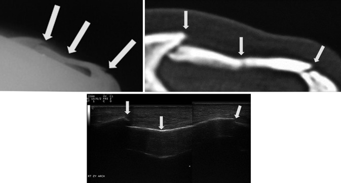
Sensitivity and specificity of USG in assessing zygomatic arch fractures were 100% and 100%, respectively; and that of mandibular fractures were 94.74% and 100%, respectively. Overall sensitivity, specificity, positive predictive value, and negative predictive value of USG against CT in diagnosing zygomatic arch and mandibular fractures were found out to be 97.4%, 100%, 100%, and 66.7%, respectively.
USG is a very reliable tool in detection of fractures involving zygomatic arch and mandible. It can be used for screening of suspected fractures of zygomatic arch and mandible to avoid unnecessary radiation exposure from conventional radiography and CT scans.
以下研究的目的是评估超声检查(USG)与传统放射照相术和计算机断层扫描(CT)在诊断颧弓和下颌骨骨折方面的效用。
本研究共纳入40例疑似颧弓骨折和/或下颌骨骨折的患者。将患者分为两组(一组为颧弓骨折,另一组为下颌骨骨折),每组20例。使用频率为10MHz的小型线性探头(LA435,西门子Acuson Antares)进行超声检查。将CT和传统放射照相的数据与超声检查的数据进行比较。
超声检查评估颧弓骨折的敏感性和特异性分别为100%和100%;评估下颌骨骨折的敏感性和特异性分别为94.74%和100%。超声检查相对于CT诊断颧弓和下颌骨骨折的总体敏感性、特异性、阳性预测值和阴性预测值分别为97.4%、100%、100%和66.7%。
超声检查是检测涉及颧弓和下颌骨骨折的非常可靠的工具。它可用于筛查疑似颧弓和下颌骨骨折,以避免传统放射照相术和CT扫描带来的不必要辐射暴露。