Nusca S, Canterini S, Palladino G, Bruno F, Mangia F, Erickson R P, Fiorenza M T
Department of Psychology, Section of Neuroscience and "Daniel Bovet" Neurobiology Research Center, Sapienza University of Rome, 00185 Rome, Italy.
Department of Pediatrics, University of AZ, Tucson AZ85724-5073, USA.
Neurobiol Dis. 2014 Oct;70:117-26. doi: 10.1016/j.nbd.2014.06.012. Epub 2014 Jun 24.
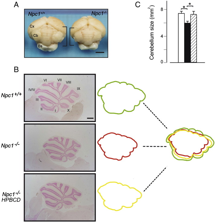
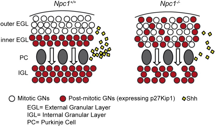
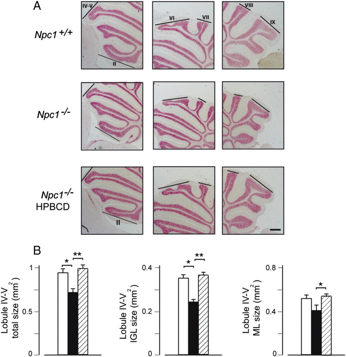
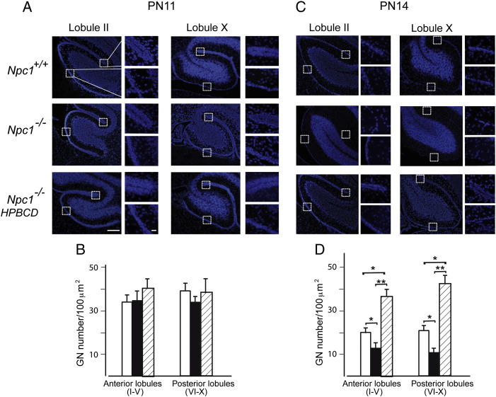
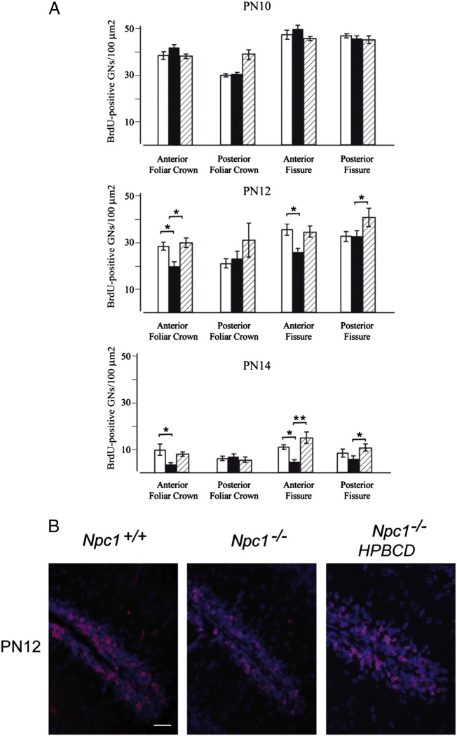
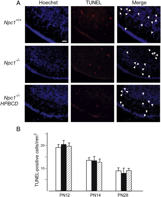
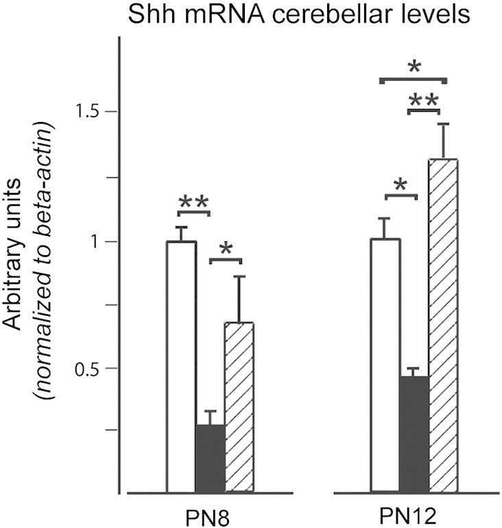
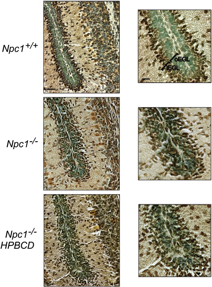
In this study we show that postnatal development of cerebellar granule neurons (GNs) is defective in Npc1(-/-) mice. Compared to age-matched wild-type littermates, there is an accelerated disappearance of the external granule layer (EGL) in these mice. This is due to a premature exit from the cell cycle of GN precursors residing at the level of the EGL. As a consequence, the size of cerebellar lobules of these mice displays a 20%-25% reduction compared to that of age-matched wild-type mice. This size reduction is detectable at post-natal day 28 (PN28), when cerebellar GN development is completed while signs of neuronal atrophy are not yet apparent. Based on the analysis of EGL thickness and the determination of proliferating GN fractions at increasing developmental times (PN8-PN14), we trace the onset of this GN developmental defect during the second postnatal week. We also show that during this developmental time Shh transcripts undergo a significant reduction in Npc1(-/-) mice compared to age-matched wild-type mice. In light of the mitogenic activity of Shh on GNs, this observation further supports the presence of defective GN proliferation in Npc1(-/-) mice. A single injection of hydroxypropyl-β-cyclodextrin at PN7 rescues this defect, restoring the normal patterns of granule neuron proliferation and cerebellar lobule size. To our knowledge, these findings identify a novel developmental defect that was underappreciated in previous studies. This defect was probably overlooked because Npc1 loss-of-function does not affect cerebellar foliation and causes the internal granule layer and molecular layer to decrease proportionally, giving rise to a normally appearing, yet harmoniously smaller, cerebellum.
在本研究中,我们发现Npc1基因敲除(Npc1-/-)小鼠的小脑颗粒神经元(GNs)出生后发育存在缺陷。与年龄匹配的野生型同窝小鼠相比,这些小鼠的外颗粒层(EGL)加速消失。这是由于位于EGL水平的GN前体细胞周期过早退出所致。因此,与年龄匹配的野生型小鼠相比,这些小鼠的小脑小叶大小减少了20%-25%。这种大小减少在出生后第28天(PN28)即可检测到,此时小脑GN发育完成,而神经元萎缩迹象尚不明显。基于对EGL厚度的分析以及在发育时间增加时(PN8-PN14)增殖GN比例的测定,我们追踪到这种GN发育缺陷在出生后第二周开始出现。我们还表明,在这个发育阶段,与年龄匹配的野生型小鼠相比,Npc1-/-小鼠中Shh转录本显著减少。鉴于Shh对GNs的促有丝分裂活性,这一观察结果进一步支持了Npc1-/-小鼠中存在有缺陷的GN增殖。在PN7单次注射羟丙基-β-环糊精可挽救这一缺陷,恢复颗粒神经元增殖和小脑小叶大小的正常模式。据我们所知,这些发现确定了一种在先前研究中未被充分认识的新型发育缺陷。这种缺陷可能被忽视了,因为Npc1功能丧失并不影响小脑叶形成,且导致内颗粒层和分子层成比例减少,从而形成外观正常但和谐缩小的小脑。