Orthopaedic and Sports Traumatology Department, Carolina Medical Center, Pory 78, 02-757, Warsaw, Poland.
Department of Descriptive and Clinical Anatomy, Medical University of Warsaw, Chalbinskiego 5, 02-004, Warsaw, Poland.
Knee Surg Sports Traumatol Arthrosc. 2015 Nov;23(11):3143-50. doi: 10.1007/s00167-014-3146-7. Epub 2014 Jun 28.
Recently, the configuration of the anterior cruciate ligament (ACL) from its direct femoral insertion to midsubstance was found to be flat. This might have an important impact for anatomical ACL reconstruction. The purpose of this anatomical study was to evaluate the macroscopic appearance of the ACL from femoral to midsubstance.
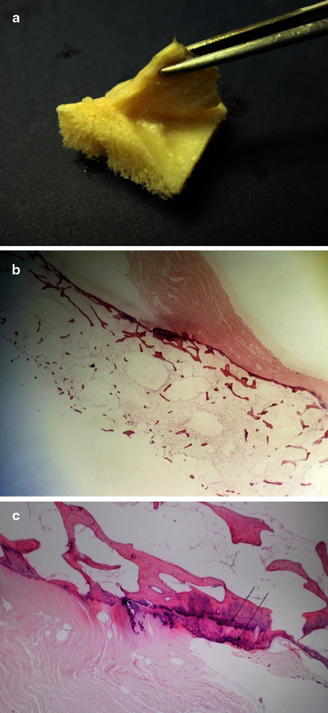
The ACL was dissected in 111 human fresh frozen cadaver knees from its femoral insertion to midsubstance, and the shape was described. The anatomical findings were documented on digital photographs and on video. Thirty knees were sent for computed tomography (CT), magnetic resonance imaging (MRI) and histology of the femoral ACL insertion.
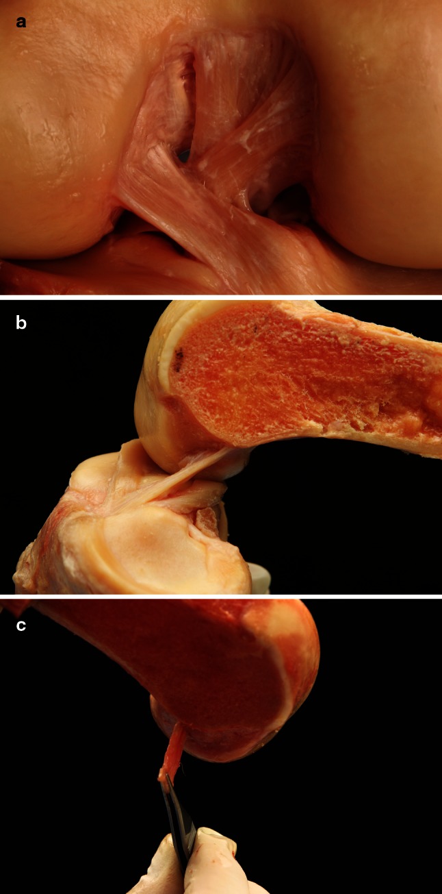
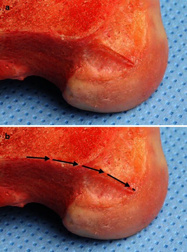
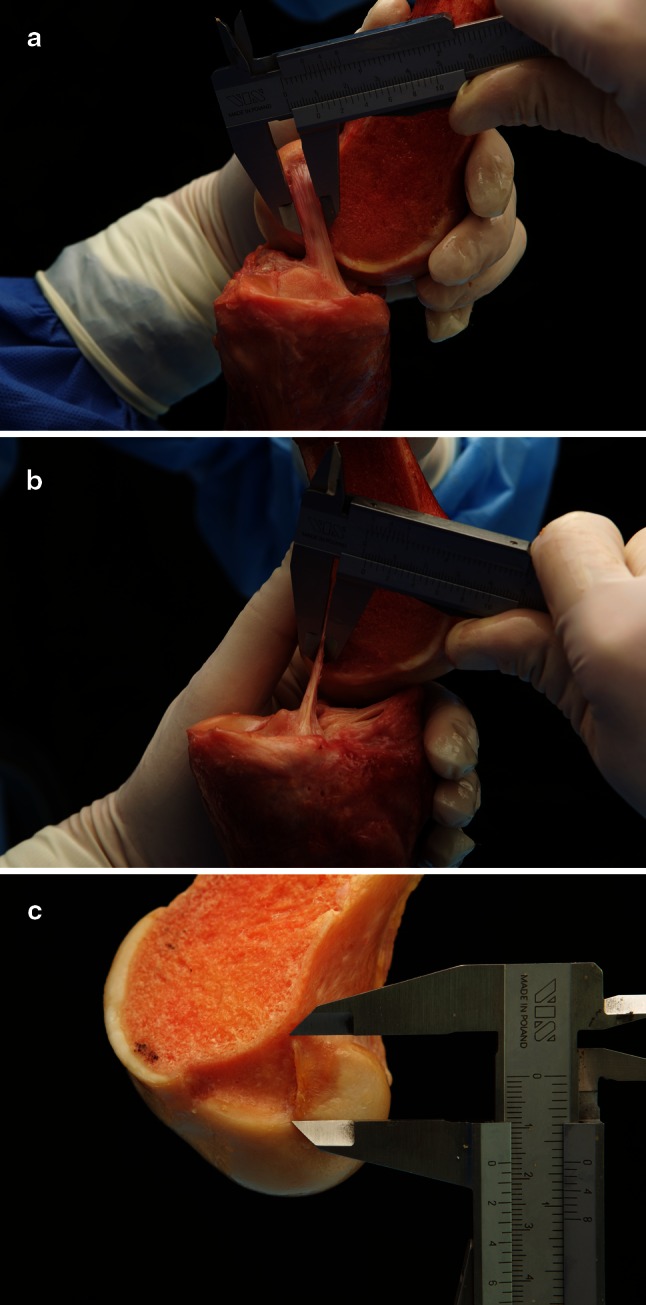
Two millimetres from its direct femoral insertion, the ACL fibres formed a flat ribbon in all dissected knees without a clear separation between AM and PL bundles. The ribbon was in exact continuity of the posterior femoral cortex. The width of the ribbon was between 11.43 and 16.18 mm and the thickness of the ACL was only 2.54-3.38 mm. 3D CT, MRI and the histological examination confirmed above findings.
This is a detailed anatomical study describing the ribbon-like structure of the ACL from its femoral insertion to midsubstance. A key point was to carefully remove the surface fibrous membrane of the ACL. A total of 2-3 mm from its bony femoral insertion, the ACL formed a flat ribbon without a clear separation between AM and PL bundles. The ribbon was in exact continuity of the posterior femoral cortex. The findings of a flat ligament may change the future approach to femoral ACL footprint and midsubstance ACL reconstruction and to graft selection.
最近发现前交叉韧带(ACL)从其直接股骨插入到中体的配置是平坦的。这可能对解剖 ACL 重建有重要影响。本解剖研究的目的是评估 ACL 从股骨到中体的大体外观。
从 111 个人体冷冻尸体膝关节的股骨插入处到中体处解剖 ACL,并描述其形状。解剖学发现记录在数字照片和视频上。30 个膝关节进行计算机断层扫描(CT)、磁共振成像(MRI)和股骨 ACL 插入的组织学检查。
在所有解剖的膝关节中,ACL 纤维在距其直接股骨插入处 2 毫米处形成扁平带,AM 和 PL 束之间没有明显的分离。该带与后股骨皮质完全连续。带的宽度在 11.43 到 16.18 毫米之间,ACL 的厚度仅为 2.54-3.38 毫米。3D CT、MRI 和组织学检查证实了上述发现。
这是一项详细的解剖研究,描述了 ACL 从股骨插入处到中体的带状结构。一个关键点是要小心地去除 ACL 的表面纤维膜。在距其骨性股骨插入处 2-3 毫米处,ACL 形成了一个没有 AM 和 PL 束之间明显分离的扁平带。该带与后股骨皮质完全连续。韧带平坦的发现可能会改变未来对股骨 ACL 足印和 ACL 中体重建以及移植物选择的方法。