Gangi Setty Thanuja, Cho Christine, Govindappa Sowmya, Apicella Michael A, Ramaswamy S
Institute for Stem Cell Biology and Regenerative Medicine, NCBS Campus, GKVK Post, Bangalore, Karnataka 560 065, India.
Department of Microbiology, Carver College of Medicine, University of Iowa, Iowa City, IA 52242-1109, USA.
Acta Crystallogr D Biol Crystallogr. 2014 Jul;70(Pt 7):1801-11. doi: 10.1107/S139900471400830X. Epub 2014 Jun 24.
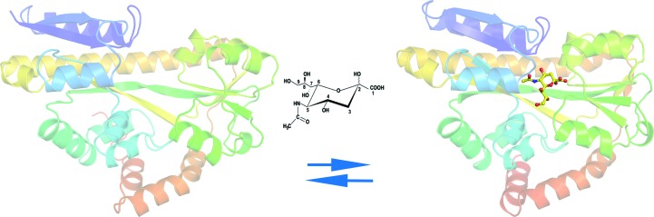
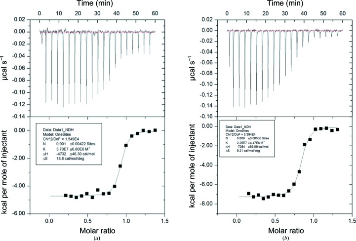
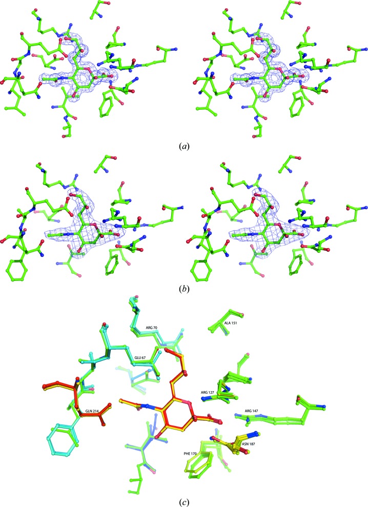
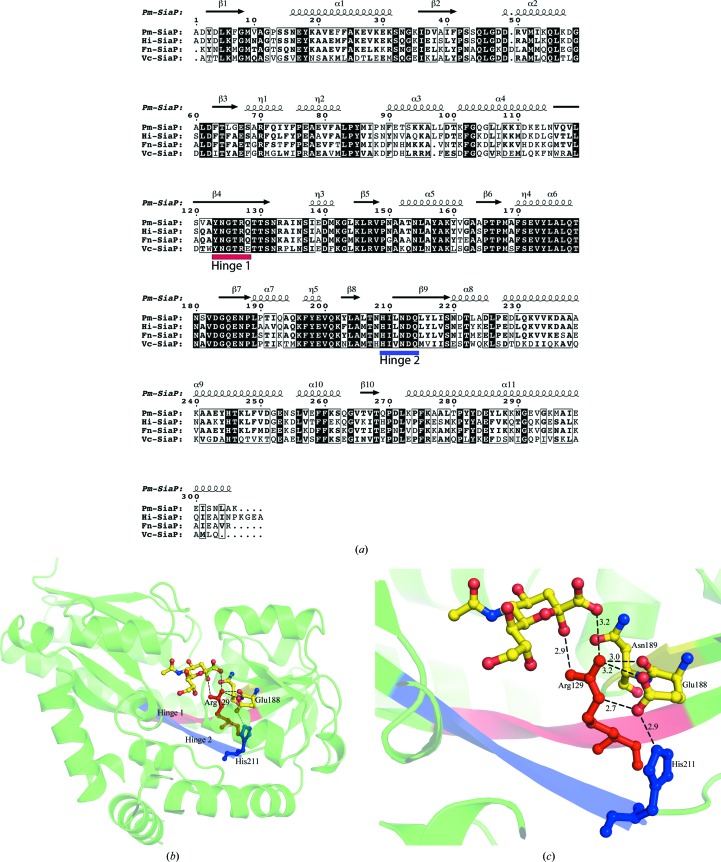
Sialic acids are a family of related nine-carbon sugar acids that play important roles in both eukaryotes and prokaryotes. These sialic acids are incorporated/decorated onto lipooligosaccharides as terminal sugars in multiple bacteria to evade the host immune system. Many pathogenic bacteria scavenge sialic acids from their host and use them for molecular mimicry. The first step of this process is the transport of sialic acid to the cytoplasm, which often takes place using a tripartite ATP-independent transport system consisting of a periplasmic binding protein and a membrane transporter. In this paper, the structural characterization of periplasmic binding proteins from the pathogenic bacteria Fusobacterium nucleatum, Pasteurella multocida and Vibrio cholerae and their thermodynamic characterization are reported. The binding affinities of several mutations in the Neu5Ac binding site of the Haemophilus influenzae protein are also reported. The structure and the thermodynamics of the binding of sugars suggest that all of these proteins have a very well conserved binding pocket and similar binding affinities. A significant conformational change occurs when these proteins bind the sugar. While the C1 carboxylate has been identified as the primary binding site, a second conserved hydrogen-bonding network is involved in the initiation and stabilization of the conformational states.
唾液酸是一类相关的九碳糖酸,在真核生物和原核生物中都发挥着重要作用。这些唾液酸作为末端糖被整合/修饰到多种细菌的脂寡糖上,以逃避宿主免疫系统。许多病原菌从宿主中获取唾液酸并将其用于分子模拟。这个过程的第一步是将唾液酸转运到细胞质中,这通常通过一个由周质结合蛋白和膜转运蛋白组成的不依赖ATP的三方转运系统来完成。本文报道了来自病原菌具核梭杆菌、多杀巴斯德菌和霍乱弧菌的周质结合蛋白的结构表征及其热力学表征。还报道了流感嗜血杆菌蛋白Neu5Ac结合位点中几个突变的结合亲和力。糖结合的结构和热力学表明,所有这些蛋白都有一个非常保守的结合口袋和相似的结合亲和力。当这些蛋白结合糖时会发生显著的构象变化。虽然C1羧酸盐已被确定为主要结合位点,但第二个保守的氢键网络参与了构象状态的起始和稳定。