Bocan Thomas M, Panchal Rekha G, Bavari Sina
Molecular and Translational Sciences, US Army Medical Research Institute of Infectious Diseases (USAMRIID), 1425 Porter Street, Ft. Detrick, MD, 21702, USA,
Mol Imaging Biol. 2015 Feb;17(1):4-17. doi: 10.1007/s11307-014-0759-7.
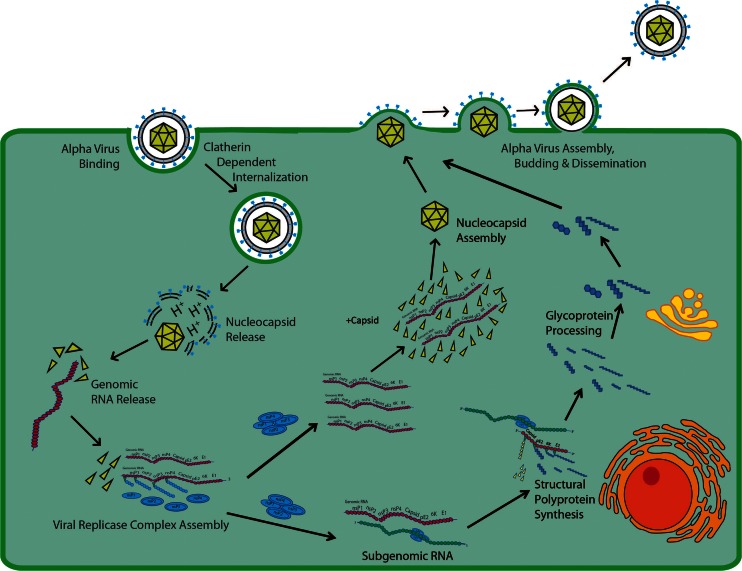
While preclinical and clinical imaging have been applied to drug discovery/development and characterization of disease pathology, few examples exist where imaging has been used to evaluate infectious agents or countermeasures to biosafety level (BSL)3/4 threat agents. Viruses engineered with reporter constructs, i.e., enzymes and receptors, which are amenable to detection by positron emission tomography (PET), single photon emission tomography (SPECT), or magnetic resonance imaging (MRI) have been used to evaluate the biodistribution of viruses containing specific therapeutic or gene transfer payloads. Bioluminescence and nuclear approaches involving engineered reporters, direct labeling of bacteria with radiotracers, or tracking bacteria through their constitutively expressed thymidine kinase have been utilized to characterize viral and bacterial pathogens post-infection. Most PET, SPECT, CT, or MRI approaches have focused on evaluating host responses to the pathogens such as inflammation, brain neurochemistry, and structural changes and on assessing the biodistribution of radiolabeled drugs. Imaging has the potential when applied preclinically to the development of countermeasures against BSL3/4 threat agents to address the following: (1) presence, biodistribution, and time course of infection in the presence or absence of drug; (2) binding of the therapeutic to the target; and (3) expression of a pharmacologic effect either related to drug mechanism, efficacy, or safety. Preclinical imaging could potentially provide real-time dynamic tools to characterize the pathogen and animal model and for developing countermeasures under the U.S. FDA Animal Rule provision with high confidence of success and clinical benefit.
虽然临床前和临床成像已应用于药物发现/开发以及疾病病理学特征分析,但利用成像来评估感染因子或针对生物安全3/4级威胁因子的应对措施的例子却很少。用报告基因构建体(即酶和受体)进行工程改造的病毒,适合通过正电子发射断层扫描(PET)、单光子发射断层扫描(SPECT)或磁共振成像(MRI)进行检测,已被用于评估含有特定治疗或基因传递载荷的病毒的生物分布。涉及工程报告基因的生物发光和核方法、用放射性示踪剂直接标记细菌或通过其组成性表达的胸苷激酶追踪细菌,已被用于表征感染后病毒和细菌病原体的特征。大多数PET、SPECT、CT或MRI方法都集中在评估宿主对病原体的反应,如炎症、脑神经化学和结构变化,以及评估放射性标记药物的生物分布。临床前应用成像技术来开发针对生物安全3/4级威胁因子的应对措施时,有潜力解决以下问题:(1)在有或没有药物的情况下感染的存在、生物分布和时间进程;(2)治疗药物与靶点的结合;(3)与药物机制、疗效或安全性相关的药理作用的表达。临床前成像有可能提供实时动态工具,以表征病原体和动物模型,并根据美国食品药品监督管理局的动物规则条款开发应对措施,从而有很高的成功和临床获益把握。