Zhang Qiu-Yue, Fu Jian-Hua, Xue Xin-Dong
Department of Pediatrics, Shengjing Hospital of China Medical University, Shenyang, Liaoning 110004, P.R. China ; Pediatrics Intensive Care Units, First Affiliated Hospital of Harbin Medical University, Harbin, Heilongjiang 150001, P.R. China.
Department of Pediatrics, Shengjing Hospital of China Medical University, Shenyang, Liaoning 110004, P.R. China.
Exp Ther Med. 2014 Aug;8(2):493-498. doi: 10.3892/etm.2014.1739. Epub 2014 May 28.
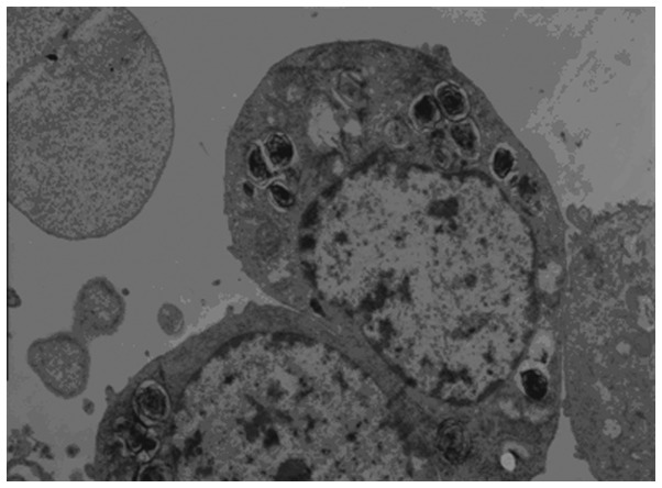
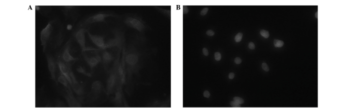
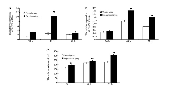
The aim of the present study was to investigate water transport dysfunction in alveolar epithelial type II cells (AECII), which were exposed to hyperoxia, and to investigate the mechanism of pulmonary edema resulting from hyperoxic lung injury. The lung cells of newborn rats were isolated for primary cell culture and divided into control and experimental groups. The control and experimental group cells were placed into a normoxic incubator (oxygen volume fraction, 0.21) or hyperoxic incubator (oxygen volume fraction, 0.9), respectively. Twenty-four, 48 and 72 h after cell attachment, the gene transcription and protein expression levels of aquaporin-1 (AQP1) were detected via quantitative polymerase chain reaction and western blot analysis. Flow cytometry was conducted to detect the volume of the cells in the experimental and control groups. In the present study, it was identified that AQP1 expression and cell volume were greater in the experimental group when compared with the control group. Thus, hyperoxia may disturb the gene expression regulation of AQP1 in AECII, resulting in water transport dysfunction. This may be one of the mechanisms underlying pulmonary edema caused by hyperoxic lung injury.
本研究的目的是调查暴露于高氧环境下的II型肺泡上皮细胞(AECII)的水转运功能障碍,并探究高氧肺损伤导致肺水肿的机制。分离新生大鼠的肺细胞进行原代细胞培养,并分为对照组和实验组。将对照组和实验组细胞分别置于常氧培养箱(氧体积分数为0.21)或高氧培养箱(氧体积分数为0.9)中。细胞贴壁后24、48和72小时,通过定量聚合酶链反应和蛋白质印迹分析检测水通道蛋白-1(AQP1)的基因转录和蛋白表达水平。进行流式细胞术检测实验组和对照组细胞的体积。在本研究中,发现与对照组相比,实验组中AQP1表达和细胞体积更大。因此,高氧可能会干扰AECII中AQP1的基因表达调控,导致水转运功能障碍。这可能是高氧肺损伤引起肺水肿的潜在机制之一。