Canales Norma Angélica Galicia, Marina Vicente Madrid, Castro Jorge Salmerón, Jiménez Alfredo Antúnez, Mendoza-Hernández Guillermo, McCARRON Elizabeth Langley, Roman Margarita Bahena, Castro-Romero Julieta Ivone
Research Center on Infection Diseases, National Institute of Public Health, Cuernavaca, Morelos 62100, Mexico.
Epidemiology and Health Services Research Unit, National Institute of Social Security, Cuernavaca, Morelos 62450, Mexico.
Oncol Lett. 2014 Aug;8(2):939-947. doi: 10.3892/ol.2014.2195. Epub 2014 May 28.
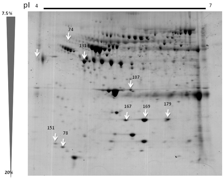
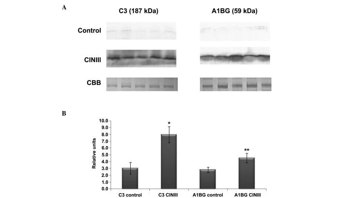
The present study aimed to analyze sera proteins in females with cervical intraepithelial neoplasia, grade III (CIN III) and in healthy control females, in order to identify a potential biomarker which detects lesions that have a greater probability of cervical transformation. The present study investigated five sera samples from females who were Human Papilloma Virus (HPV) 16 and who had been histopathologically diagnosed with CIN III, as well as five sera samples from healthy control females who were HPV-negative. Protein separation was performed using two-dimensional (2D) gel electrophoresis and the proteins were stained with Colloidal Coommassie Blue. Quantitative analysis was performed using ImageMaster 2D Platinum 6.0 software. Peptide sequence identification was performed using a nano-LC ESIMS/MS system. The proteins with the highest Mascot score were validated using western blot analysis in an additional 55 sera samples from the control and CIN III groups. The eight highest score spots that were found to be overexpressed in the CIN III sera group were identified as α-1-B glycoprotein (A1BG), complement component 3 (C3), a pro-apolipoprotein, two apolipoproteins and three haptoglobins. Only A1BG and C3 were validated using western blot analysis, and the bands were compared between the two groups using densitometry analysis. The relative density of the bands of A1BG and C3 was found to be greater in all of the serum samples from the females with CIN III, compared with those of the individuals in the control group. In summary, the present study identified two proteins whose expression was elevated in females with CIN III, suggesting that they could be used as biomarkers for CIN III. However, further investigations are required in order to assess the expression of A1BG and C3 in different pre-malignant lesions.
本研究旨在分析宫颈上皮内瘤变III级(CIN III)女性和健康对照女性的血清蛋白,以确定一种潜在的生物标志物,用于检测具有较高宫颈癌变可能性的病变。本研究调查了5例感染人乳头瘤病毒(HPV)16且经组织病理学诊断为CIN III的女性血清样本,以及5例HPV阴性的健康对照女性血清样本。采用二维(2D)凝胶电泳进行蛋白质分离,并用考马斯亮蓝胶体染色蛋白质。使用ImageMaster 2D Platinum 6.0软件进行定量分析。使用纳升液相色谱电喷雾串联质谱系统进行肽序列鉴定。在另外55例来自对照组和CIN III组的血清样本中,使用蛋白质免疫印迹分析对Mascot得分最高的蛋白质进行验证。在CIN III血清组中发现过表达的8个得分最高的斑点被鉴定为α-1-B糖蛋白(A1BG)、补体成分3(C3)、一种载脂蛋白前体、两种载脂蛋白和三种触珠蛋白。仅使用蛋白质免疫印迹分析对A1BG和C3进行验证,并使用光密度分析比较两组之间的条带。与对照组个体相比,在所有CIN III女性的血清样本中,A1BG和C3条带的相对密度更高。总之,本研究鉴定出两种在CIN III女性中表达升高的蛋白质,表明它们可作为CIN III的生物标志物。然而,需要进一步研究以评估A1BG和C3在不同癌前病变中的表达。