• 文献检索
  • 文档翻译
  • 深度研究
  • 学术资讯
  • Suppr Zotero 插件Zotero 插件
  • 邀请有礼
  • 套餐&价格
  • 历史记录
应用&插件
Suppr Zotero 插件Zotero 插件浏览器插件Mac 客户端Windows 客户端微信小程序
定价
高级版会员购买积分包购买API积分包
服务
文献检索文档翻译深度研究API 文档MCP 服务
关于我们
关于 Suppr公司介绍联系我们用户协议隐私条款
关注我们

Suppr 超能文献

核心技术专利:CN118964589B侵权必究
粤ICP备2023148730 号-1Suppr @ 2026

文献检索

告别复杂PubMed语法,用中文像聊天一样搜索,搜遍4000万医学文献。AI智能推荐,让科研检索更轻松。

立即免费搜索

文件翻译

保留排版,准确专业,支持PDF/Word/PPT等文件格式,支持 12+语言互译。

免费翻译文档

深度研究

AI帮你快速写综述,25分钟生成高质量综述,智能提取关键信息,辅助科研写作。

立即免费体验

基于单受试者的全脑脑磁图慢波成像方法用于检测轻度创伤性脑损伤患者的异常情况。

Single-subject-based whole-brain MEG slow-wave imaging approach for detecting abnormality in patients with mild traumatic brain injury.

作者信息

Huang Ming-Xiong, Nichols Sharon, Baker Dewleen G, Robb Ashley, Angeles Annemarie, Yurgil Kate A, Drake Angela, Levy Michael, Song Tao, McLay Robert, Theilmann Rebecca J, Diwakar Mithun, Risbrough Victoria B, Ji Zhengwei, Huang Charles W, Chang Douglas G, Harrington Deborah L, Muzzatti Laura, Canive Jose M, Christopher Edgar J, Chen Yu-Han, Lee Roland R

机构信息

Radiology, Research, and Psychiatry Services, VA San Diego Healthcare System, San Diego, CA, USA ; Department of Radiology, University of California, San Diego, CA, USA.

Department of Neuroscience, University of California, San Diego, CA, USA.

出版信息

Neuroimage Clin. 2014 Jun 16;5:109-19. doi: 10.1016/j.nicl.2014.06.004. eCollection 2014.

DOI:10.1016/j.nicl.2014.06.004
PMID:25009772
原文链接:
https://pmc.ncbi.nlm.nih.gov/articles/PMC4087185/
Abstract

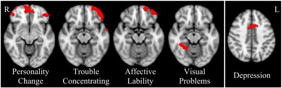
Traumatic brain injury (TBI) is a leading cause of sustained impairment in military and civilian populations. However, mild TBI (mTBI) can be difficult to detect using conventional MRI or CT. Injured brain tissues in mTBI patients generate abnormal slow-waves (1-4 Hz) that can be measured and localized by resting-state magnetoencephalography (MEG). In this study, we develop a voxel-based whole-brain MEG slow-wave imaging approach for detecting abnormality in patients with mTBI on a single-subject basis. A normative database of resting-state MEG source magnitude images (1-4 Hz) from 79 healthy control subjects was established for all brain voxels. The high-resolution MEG source magnitude images were obtained by our recent Fast-VESTAL method. In 84 mTBI patients with persistent post-concussive symptoms (36 from blasts, and 48 from non-blast causes), our method detected abnormalities at the positive detection rates of 84.5%, 86.1%, and 83.3% for the combined (blast-induced plus with non-blast causes), blast, and non-blast mTBI groups, respectively. We found that prefrontal, posterior parietal, inferior temporal, hippocampus, and cerebella areas were particularly vulnerable to head trauma. The result also showed that MEG slow-wave generation in prefrontal areas positively correlated with personality change, trouble concentrating, affective lability, and depression symptoms. Discussion is provided regarding the neuronal mechanisms of MEG slow-wave generation due to deafferentation caused by axonal injury and/or blockages/limitations of cholinergic transmission in TBI. This study provides an effective way for using MEG slow-wave source imaging to localize affected areas and supports MEG as a tool for assisting the diagnosis of mTBI.

摘要

创伤性脑损伤(TBI)是导致军人和 civilian populations 持续受损的主要原因。然而,使用传统的MRI或CT很难检测出轻度TBI(mTBI)。mTBI患者受损的脑组织会产生异常慢波(1-4Hz),可通过静息态脑磁图(MEG)进行测量和定位。在本研究中,我们开发了一种基于体素的全脑MEG慢波成像方法,用于在单个体素基础上检测mTBI患者的异常情况。为所有脑体素建立了一个由79名健康对照受试者的静息态MEG源强度图像(1-4Hz)组成的标准数据库。高分辨率MEG源强度图像通过我们最近的Fast-VESTAL方法获得。在84名有持续脑震荡后症状的mTBI患者中(36名由爆炸引起,48名由非爆炸原因引起),我们的方法在联合组(爆炸引起加非爆炸原因)、爆炸组和非爆炸mTBI组中的阳性检测率分别为84.5%、86.1%和83.3%。我们发现前额叶、顶叶后部、颞下叶、海马体和小脑区域特别容易受到头部创伤。结果还表明,前额叶区域的MEG慢波产生与人格改变、注意力不集中、情感不稳定和抑郁症状呈正相关。讨论了TBI中由于轴突损伤和/或胆碱能传递受阻/受限导致的去传入作用而产生MEG慢波的神经元机制。本研究为使用MEG慢波源成像定位受影响区域提供了一种有效方法,并支持将MEG作为辅助诊断mTBI的工具。

https://cdn.ncbi.nlm.nih.gov/pmc/blobs/7472/4087185/21c4c233e1b5/gr5.jpg
https://cdn.ncbi.nlm.nih.gov/pmc/blobs/7472/4087185/2296216b3d8a/gr1.jpg
https://cdn.ncbi.nlm.nih.gov/pmc/blobs/7472/4087185/7a2aa915624f/gr2.jpg
https://cdn.ncbi.nlm.nih.gov/pmc/blobs/7472/4087185/73792271ffc1/gr3.jpg
https://cdn.ncbi.nlm.nih.gov/pmc/blobs/7472/4087185/47fa0fa5b8ba/gr4.jpg
https://cdn.ncbi.nlm.nih.gov/pmc/blobs/7472/4087185/21c4c233e1b5/gr5.jpg

相似文献

1
Single-subject-based whole-brain MEG slow-wave imaging approach for detecting abnormality in patients with mild traumatic brain injury.基于单受试者的全脑脑磁图慢波成像方法用于检测轻度创伤性脑损伤患者的异常情况。
Neuroimage Clin. 2014 Jun 16;5:109-19. doi: 10.1016/j.nicl.2014.06.004. eCollection 2014.
2
An automatic MEG low-frequency source imaging approach for detecting injuries in mild and moderate TBI patients with blast and non-blast causes.一种用于检测因爆炸和非爆炸原因导致的轻度和中度创伤性脑损伤患者损伤的自动 MEG 低频源成像方法。
Neuroimage. 2012 Jul 16;61(4):1067-82. doi: 10.1016/j.neuroimage.2012.04.029. Epub 2012 Apr 20.
3
The role of biomarkers and MEG-based imaging markers in the diagnosis of post-traumatic stress disorder and blast-induced mild traumatic brain injury.生物标志物和基于脑磁图的成像标志物在创伤后应激障碍和爆炸所致轻度创伤性脑损伤诊断中的作用。
Psychoneuroendocrinology. 2016 Jan;63:398-409. doi: 10.1016/j.psyneuen.2015.02.008. Epub 2015 Feb 23.
4
Integrated imaging approach with MEG and DTI to detect mild traumatic brain injury in military and civilian patients.采用 MEG 和 DTI 的综合成像方法检测军事和平民患者的轻度创伤性脑损伤。
J Neurotrauma. 2009 Aug;26(8):1213-26. doi: 10.1089/neu.2008.0672.
5
Resting-State Magnetoencephalography Reveals Different Patterns of Aberrant Functional Connectivity in Combat-Related Mild Traumatic Brain Injury.静息态脑磁图揭示了与战斗相关的轻度创伤性脑损伤中异常功能连接的不同模式。
J Neurotrauma. 2017 Apr 1;34(7):1412-1426. doi: 10.1089/neu.2016.4581. Epub 2016 Dec 2.
6
[Mild traumatic brain injury and postconcussive syndrome: a re-emergent questioning].[轻度创伤性脑损伤与脑震荡后综合征:一个重新出现的问题探讨]
Encephale. 2012 Sep;38(4):329-35. doi: 10.1016/j.encep.2011.07.003. Epub 2011 Aug 31.
7
Magnetoencephalography Slow-Wave Detection in Patients with Mild Traumatic Brain Injury and Ongoing Symptoms Correlated with Long-Term Neuropsychological Outcome.轻度创伤性脑损伤且伴有持续症状患者的脑磁图慢波检测与长期神经心理学结果的相关性
J Neurotrauma. 2015 Oct 1;32(19):1510-21. doi: 10.1089/neu.2014.3654. Epub 2015 Jun 18.
8
Objective documentation of traumatic brain injury subsequent to mild head trauma: multimodal brain imaging with MEG, SPECT, and MRI.轻度头部创伤后创伤性脑损伤的客观记录:采用脑磁图(MEG)、单光子发射计算机断层扫描(SPECT)和磁共振成像(MRI)的多模态脑成像
J Head Trauma Rehabil. 2007 May-Jun;22(3):141-55. doi: 10.1097/01.HTR.0000271115.29954.27.
9
Magnetoencephalography in the diagnosis of concussion.脑磁图在脑震荡诊断中的应用
Prog Neurol Surg. 2014;28:94-111. doi: 10.1159/000358768. Epub 2014 Jun 6.
10
A multisite study of the relationships between blast exposures and symptom reporting in a post-deployment active duty military population with mild traumatic brain injury.一项关于部署后现役军人轻度创伤性脑损伤群体中爆炸暴露与症状报告之间关系的多中心研究。
J Neurotrauma. 2014 Dec 1;31(23):1899-906. doi: 10.1089/neu.2014.3455. Epub 2014 Oct 24.

引用本文的文献

1
Detecting mild traumatic brain injury with MEG scan data: One-vs-K-sample tests.利用脑磁图扫描数据检测轻度创伤性脑损伤:一对多样本检验。
Imaging Neurosci (Camb). 2025 Sep 8;3. doi: 10.1162/IMAG.a.137. eCollection 2025.
2
Retraining Dorsal Visual Pathways Improves Cognitive Skills After a Mild Traumatic Brain Injury.轻度创伤性脑损伤后,重新训练背侧视觉通路可改善认知技能。
J Clin Med. 2025 Mar 26;14(7):2273. doi: 10.3390/jcm14072273.
3
Network effects of traumatic brain injury: from infra slow to high frequency oscillations and seizures.

本文引用的文献

1
MEG source imaging method using fast L1 minimum-norm and its applications to signals with brain noise and human resting-state source amplitude images.使用快速 L1 最小范数的 MEG 源成像方法及其在具有脑噪声和人类静息状态源幅度图像的信号中的应用。
Neuroimage. 2014 Jan 1;84:585-604. doi: 10.1016/j.neuroimage.2013.09.022. Epub 2013 Sep 19.
2
Complexity analysis of resting state magnetoencephalography activity in traumatic brain injury patients.创伤性脑损伤患者静息态脑磁图活动的复杂性分析。
J Neurotrauma. 2013 Oct 15;30(20):1702-9. doi: 10.1089/neu.2012.2679. Epub 2013 Aug 31.
3
Resting state magnetoencephalography functional connectivity in traumatic brain injury.
创伤性脑损伤的网络效应:从低频振荡到高频振荡及癫痫发作
J Comput Neurosci. 2025 Feb 28. doi: 10.1007/s10827-025-00895-5.
4
Neuroimaging findings and balance problems after mild traumatic brain injury: A systematic review protocol.轻度创伤性脑损伤后的神经影像学表现与平衡问题:一项系统评价方案
PLoS One. 2025 Feb 5;20(2):e0307339. doi: 10.1371/journal.pone.0307339. eCollection 2025.
5
Delta Wave MRI: fMRI of Electrophysiologic Activity.δ波磁共振成像:电生理活动的功能磁共振成像
AJNR Am J Neuroradiol. 2025 Jun 3;46(6):1203-1207. doi: 10.3174/ajnr.A8618.
6
Repetitive subconcussion results in disrupted neural activity independent of concussion history.重复性轻度脑震荡会导致神经活动紊乱,且与脑震荡病史无关。
Brain Commun. 2024 Oct 8;6(5):fcae348. doi: 10.1093/braincomms/fcae348. eCollection 2024.
7
Reduced Cortical Excitability is Associated with Cognitive Symptoms in Concussed Adolescent Football Players.皮质兴奋性降低与青少年脑震荡足球运动员的认知症状有关。
medRxiv. 2024 Nov 1:2024.09.23.24314232. doi: 10.1101/2024.09.23.24314232.
8
EMG-projected MEG high-resolution source imaging of human motor execution: Brain-muscle coupling above movement frequencies.人类运动执行的肌电图预测脑磁图高分辨率源成像:高于运动频率的脑-肌肉耦合
Imaging Neurosci (Camb). 2024 Jan 9;2:1-20. doi: 10.1162/imag_a_00056. eCollection 2024 Jan 1.
9
Magnetoencephalography for the pediatric population, indications, acquisition and interpretation for the clinician.儿科人群的脑磁图:临床医生的适应症、采集与解读
Neuroradiol J. 2025 Feb;38(1):7-20. doi: 10.1177/19714009241260801. Epub 2024 Jun 12.
10
Use of magnetic source imaging to assess recovery after severe traumatic brain injury-an MEG pilot study.使用磁源成像评估重度创伤性脑损伤后的恢复情况——一项脑磁图初步研究
Front Neurol. 2023 Nov 3;14:1257886. doi: 10.3389/fneur.2023.1257886. eCollection 2023.
创伤性脑损伤的静息态脑磁图功能连接。
J Neurosurg. 2013 Jun;118(6):1306-16. doi: 10.3171/2013.3.JNS12398. Epub 2013 Apr 19.
4
Biomarkers for the diagnosis and prognosis of mild traumatic brain injury/concussion.用于轻度创伤性脑损伤/脑震荡诊断和预后的生物标志物。
J Neurotrauma. 2013 Apr 15;30(8):657-70. doi: 10.1089/neu.2012.2439.
5
An automatic MEG low-frequency source imaging approach for detecting injuries in mild and moderate TBI patients with blast and non-blast causes.一种用于检测因爆炸和非爆炸原因导致的轻度和中度创伤性脑损伤患者损伤的自动 MEG 低频源成像方法。
Neuroimage. 2012 Jul 16;61(4):1067-82. doi: 10.1016/j.neuroimage.2012.04.029. Epub 2012 Apr 20.
6
Depression following traumatic brain injury: epidemiology, risk factors and management.创伤性脑损伤后抑郁:流行病学、危险因素和管理。
CNS Drugs. 2012 Feb 1;26(2):111-21. doi: 10.2165/11599560-000000000-00000.
7
Diffuse and spatially variable white matter disruptions are associated with blast-related mild traumatic brain injury.弥漫性和空间变异性的脑白质损伤与爆炸相关的轻度创伤性脑损伤有关。
Neuroimage. 2012 Feb 1;59(3):2017-24. doi: 10.1016/j.neuroimage.2011.10.050. Epub 2011 Oct 20.
8
Meta-analysis of facial affect recognition difficulties after traumatic brain injury.创伤性脑损伤后面部情感识别困难的荟萃分析。
Neuropsychology. 2011 May;25(3):277-285. doi: 10.1037/a0021908.
9
Blast exposure in rats with body shielding is characterized primarily by diffuse axonal injury.有身体防护的大鼠的爆炸暴露主要表现为弥漫性轴索损伤。
J Neurotrauma. 2011 Jun;28(6):947-59. doi: 10.1089/neu.2010.1540.
10
Blast-induced mild traumatic brain injury.爆炸所致轻度创伤性脑损伤。
Psychiatr Clin North Am. 2010 Dec;33(4):757-81. doi: 10.1016/j.psc.2010.08.001. Epub 2010 Oct 15.