Lemasters John J
Center for Cell Death, Injury & Regeneration, Department of Drug Discovery & Biomedical Sciences, Medical University of South Carolina, DD504 Drug Discovery Building, 70 President Street, MSC 140, Charleston, SC 29425, United States of America ; Department of Biochemistry & Molecular Biology, Medical University of South Carolina, SC, United States of America ; Institute of Theoretical & Experimental Biophysics, Russian Academy of Sciences, Pushchino, Russian Federation.
Redox Biol. 2014 Jun 12;2:749-54. doi: 10.1016/j.redox.2014.06.004. eCollection 2014.
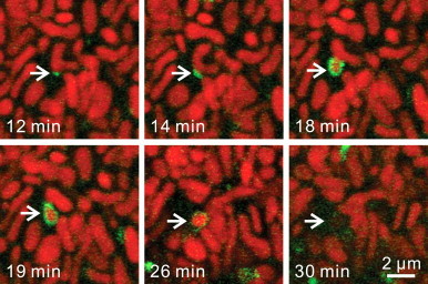
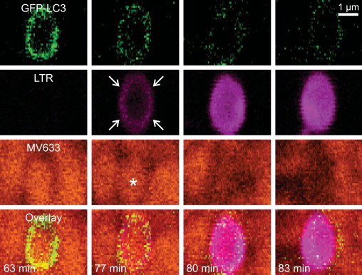
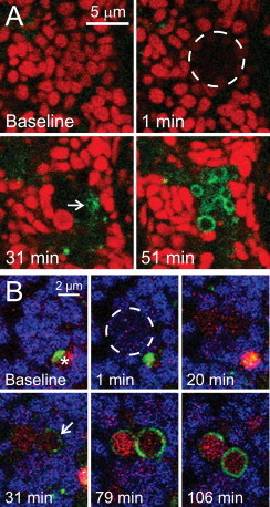
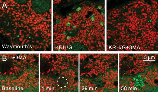
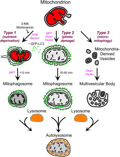
Mitophagy (mitochondrial autophagy), which removes damaged, effete and superfluous mitochondria, has several distinct variants. In Type 1 mitophagy occurring during nutrient deprivation, preautophagic structures (PAS) grow into cup-shaped phagophores that surround and sequester individual mitochondria into mitophagosomes, a process requiring phosphatidylinositol-3-kinase (PI3K) and often occurring in coordination with mitochondrial fission. After sequestration, the outer compartment of the mitophagosome acidifies, followed by mitochondrial depolarization and ultimately hydrolytic digestion in lysosomes. Mitochondrial damage stimulates Type 2 mitophagy. After photodamage to single mitochondria, depolarization occurs followed by decoration and then coalescence of autophagic LC3-containing structures on mitochondrial surfaces. Vesicular acidification then occurs. By contrast to Type 1 mitophagy, PI3K inhibition does not block Type 2 mitophagy. Further, Type 2 mitophagy is not associated with phagophore formation or mitochondrial fission. A third form of self-eating of mitochondria is formation of mitochondria-derived vesicles (MDVs) enriched in oxidized mitochondrial proteins that bud off and transit into multivesicular bodies. Topologically, the internalization of MDV by invagination of the surface of multivesicular bodies followed by vesicle scission into the lumen is a form of microautophagy, or micromitophagy (Type 3 mitophagy). Cell biological distinctions are the basis for these three types of mitophagy. Future studies are needed to better characterize the molecular and biochemical differences between Types 1, 2 and 3 mitophagy.
线粒体自噬(即线粒体的自噬作用)可清除受损、衰老及多余的线粒体,它有几种不同的形式。在营养缺乏时发生的1型线粒体自噬中,自噬前体结构(PAS)会长成杯状吞噬泡,将单个线粒体包围并隔离到线粒体自噬体中,这一过程需要磷脂酰肌醇-3-激酶(PI3K),且常与线粒体分裂协同发生。隔离后,线粒体自噬体的外腔酸化,随后线粒体去极化,最终在溶酶体中进行水解消化。线粒体损伤会刺激2型线粒体自噬。单个线粒体受到光损伤后,会发生去极化,随后自噬相关的含LC3结构会在其表面修饰并聚集,接着发生囊泡酸化。与1型线粒体自噬不同,PI3K抑制不会阻断2型线粒体自噬。此外,2型线粒体自噬与吞噬泡形成或线粒体分裂无关。线粒体自噬的第三种形式是形成富含氧化线粒体蛋白的线粒体衍生囊泡(MDV),这些囊泡会从线粒体上芽生并转运到多囊泡体中。从拓扑学角度来看,多囊泡体表面内陷使MDV内化,随后囊泡断裂进入腔内,这是一种微自噬形式,即微线粒体自噬(3型线粒体自噬)。细胞生物学差异是这三种线粒体自噬类型的基础。未来需要开展更多研究,以更好地描述1型、2型和3型线粒体自噬之间的分子和生化差异。