Boczek Tomasz, Lisek Malwina, Ferenc Bozena, Kowalski Antoni, Stepinski Dariusz, Wiktorska Magdalena, Zylinska Ludmila
Department of Molecular Neurochemistry, Medical University, Lodz, Poland.
Department of Molecular Neurochemistry, Medical University, Lodz, Poland; Department of Molecular Biology and Genetics, Aarhus University, Aarhus, Denmark.
PLoS One. 2014 Jul 11;9(7):e102352. doi: 10.1371/journal.pone.0102352. eCollection 2014.
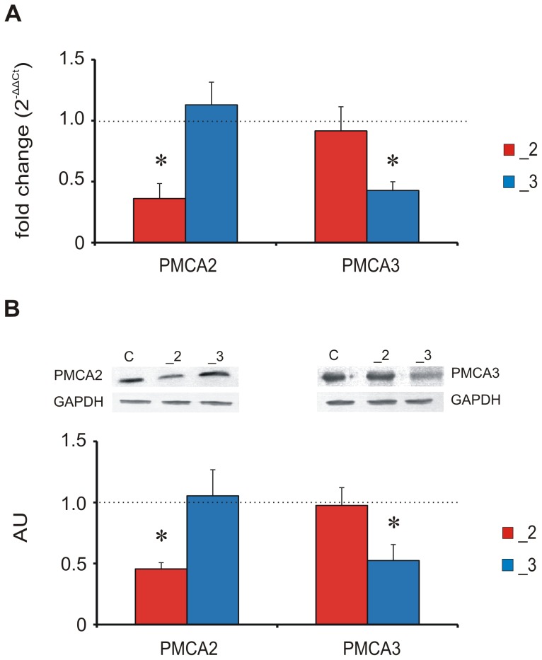
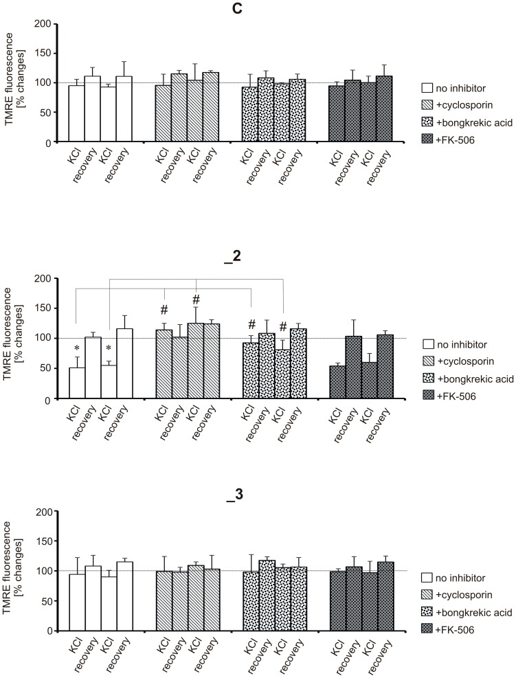
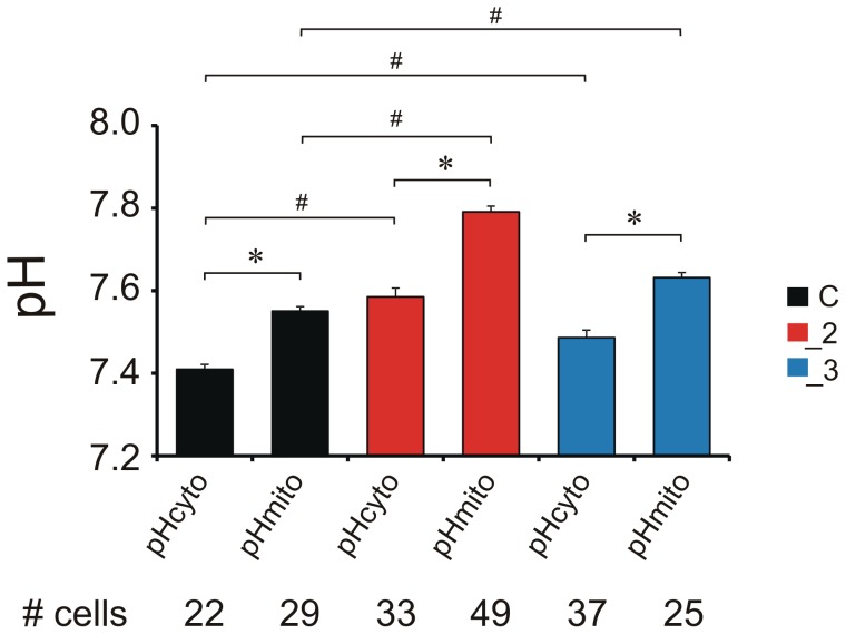
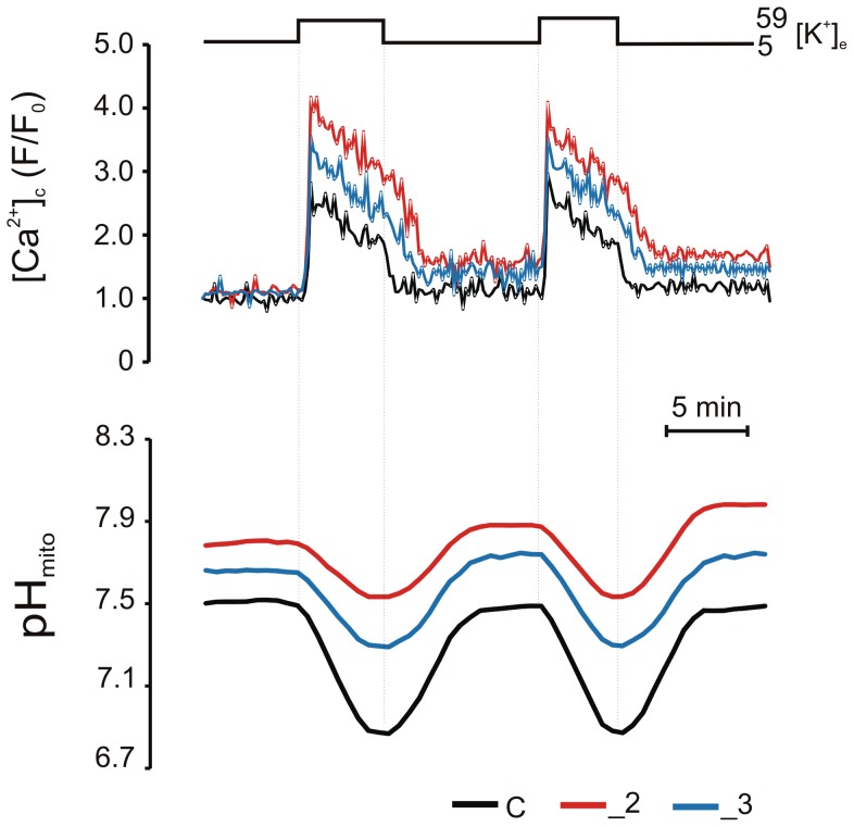
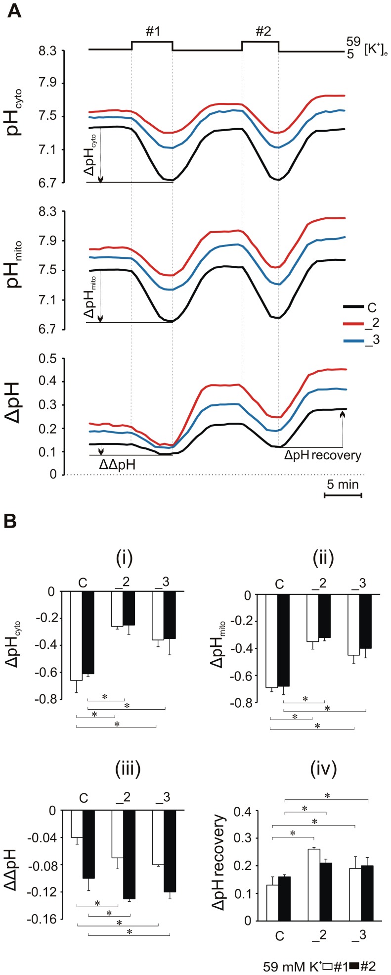
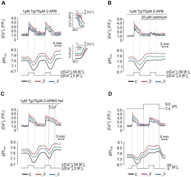
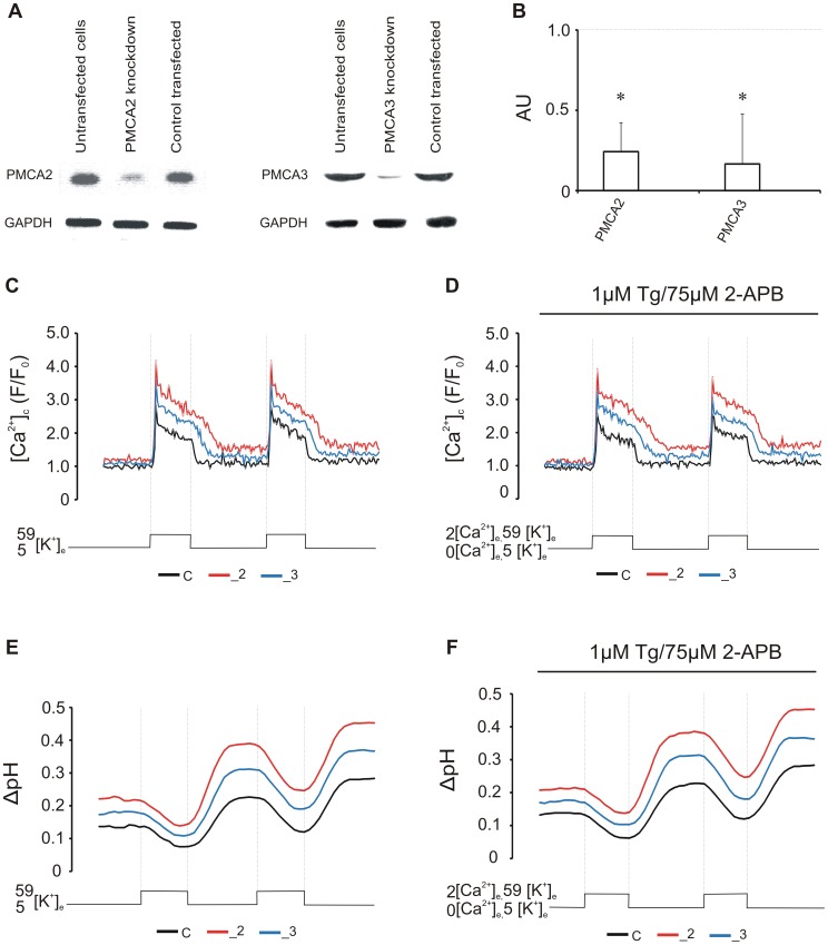
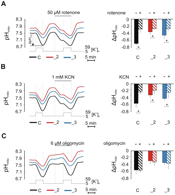
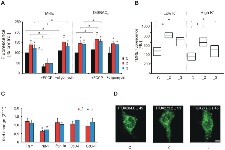
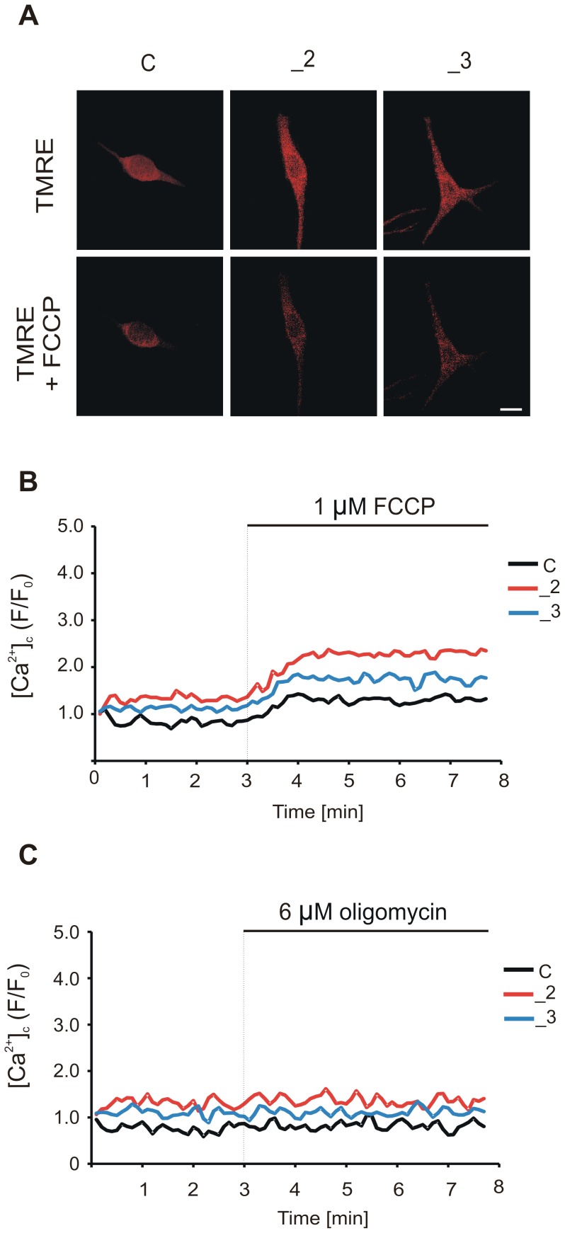
Plasma membrane Ca(2+)-ATPase (PMCA) by extruding Ca(2+) outside the cell, actively participates in the regulation of intracellular Ca(2+) concentration. Acting as Ca(2+)/H(+) counter-transporter, PMCA transports large quantities of protons which may affect organellar pH homeostasis. PMCA exists in four isoforms (PMCA1-4) but only PMCA2 and PMCA3, due to their unique localization and features, perform more specialized function. Using differentiated PC12 cells we assessed the role of PMCA2 and PMCA3 in the regulation of intracellular pH in steady-state conditions and during Ca(2+) overload evoked by 59 mM KCl. We observed that manipulation in PMCA expression elevated pHmito and pHcyto but only in PMCA2-downregulated cells higher mitochondrial pH gradient (ΔpH) was found in steady-state conditions. Our data also demonstrated that PMCA2 or PMCA3 knock-down delayed Ca(2+) clearance and partially attenuated cellular acidification during KCl-stimulated Ca(2+) influx. Because SERCA and NCX modulated cellular pH response in neglectable manner, and all conditions used to inhibit PMCA prevented KCl-induced pH drop, we considered PMCA2 and PMCA3 as mainly responsible for transport of protons to intracellular milieu. In steady-state conditions, higher TMRE uptake in PMCA2-knockdown line was driven by plasma membrane potential (Ψp). Nonetheless, mitochondrial membrane potential (Ψm) in this line was dissipated during Ca(2+) overload. Cyclosporin and bongkrekic acid prevented Ψm loss suggesting the involvement of Ca(2+)-driven opening of mitochondrial permeability transition pore as putative underlying mechanism. The findings presented here demonstrate a crucial role of PMCA2 and PMCA3 in regulation of cellular pH and indicate PMCA membrane composition important for preservation of electrochemical gradient.
质膜钙ATP酶(PMCA)通过将钙离子排出细胞外,积极参与细胞内钙离子浓度的调节。作为钙/氢反向转运体,PMCA运输大量质子,这可能会影响细胞器的pH稳态。PMCA存在四种亚型(PMCA1 - 4),但只有PMCA2和PMCA3因其独特的定位和特性而执行更特殊的功能。我们使用分化的PC12细胞评估了PMCA2和PMCA3在稳态条件下以及59 mM氯化钾诱发的钙离子过载期间对细胞内pH调节的作用。我们观察到,对PMCA表达的操控提高了线粒体pH值(pHmito)和细胞质pH值(pHcyto),但仅在PMCA2下调的细胞中,在稳态条件下发现了更高的线粒体pH梯度(ΔpH)。我们的数据还表明,在氯化钾刺激的钙离子内流期间,敲低PMCA2或PMCA3会延迟钙离子清除并部分减弱细胞酸化。由于肌浆网钙ATP酶(SERCA)和钠钙交换体(NCX)对细胞pH反应的调节作用可忽略不计,并且所有用于抑制PMCA的条件都能防止氯化钾诱导的pH下降,我们认为PMCA2和PMCA3主要负责将质子转运到细胞内环境。在稳态条件下,PMCA2敲低细胞系中更高的四甲基罗丹明乙酯(TMRE)摄取是由质膜电位(Ψp)驱动的。尽管如此,在钙离子过载期间,该细胞系中的线粒体膜电位(Ψm)会消散。环孢菌素和硼酸可防止Ψm丧失,这表明钙离子驱动的线粒体通透性转换孔开放可能是潜在的机制。此处呈现的研究结果证明了PMCA2和PMCA3在细胞pH调节中的关键作用,并表明PMCA膜组成对于维持电化学梯度很重要。