Symonds Erin L, Peiris Madusha, Page Amanda J, Chia Bridgette, Dogra Harween, Masding Abigail, Galanakis Vasileios, Atiba Michael, Bulmer David, Young Richard L, Blackshaw L Ashley
Nerve-Gut Research Laboratory, Hanson Institute, Royal Adelaide Hospital, Adelaide, South Australia, Australia.
Wingate Institute of Neurogastroenterology, Blizard Institute, Barts and The London School of Medicine & Dentistry, Queen Mary, University of London, London, UK.
Gut. 2015 Apr;64(4):618-26. doi: 10.1136/gutjnl-2014-306834. Epub 2014 Jul 11.
Inhibition of food intake and glucose homeostasis are both promoted when nutrients stimulate enteroendocrine cells (EEC) to release gut hormones. Several specific nutrient receptors may be located on EEC that respond to dietary sugars, amino acids and fatty acids. Bypass surgery for obesity and type II diabetes works by shunting nutrients to the distal gut, where it increases activation of nutrient receptors and mediator release, but cellular mechanisms of activation are largely unknown. We determined which nutrient receptors are expressed in which gut regions and in which cells in mouse and human, how they are associated with different types of EEC, how they are activated leading to hormone and 5-HT release.
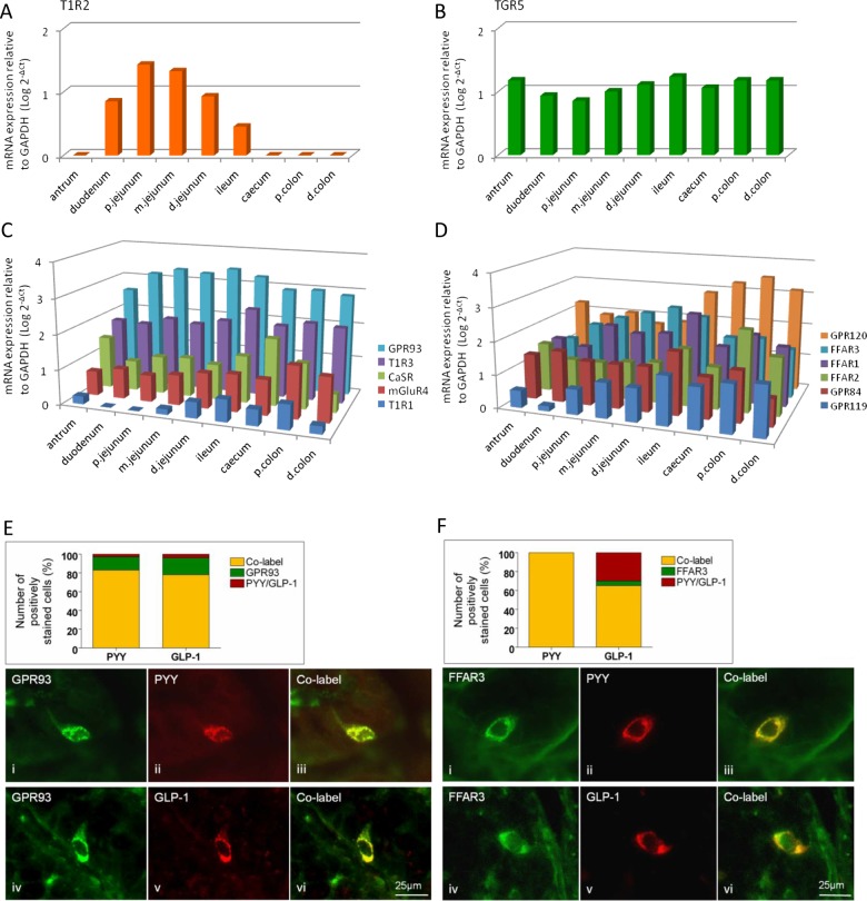
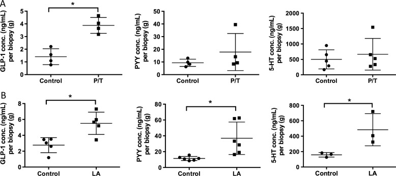
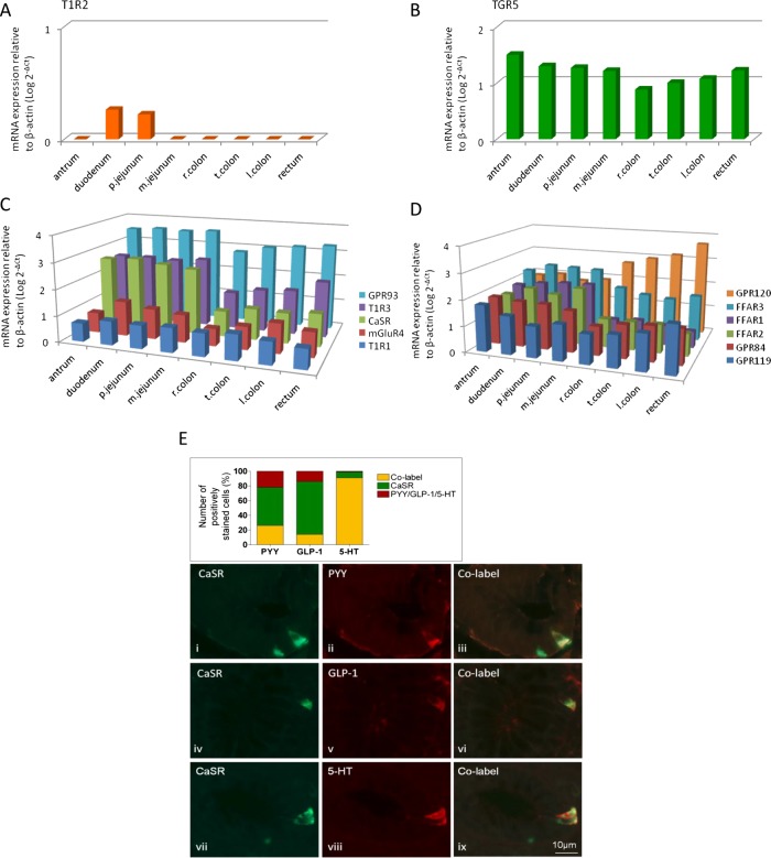
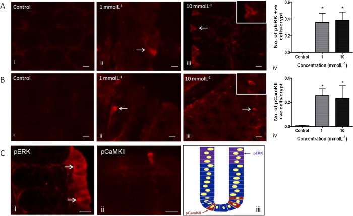
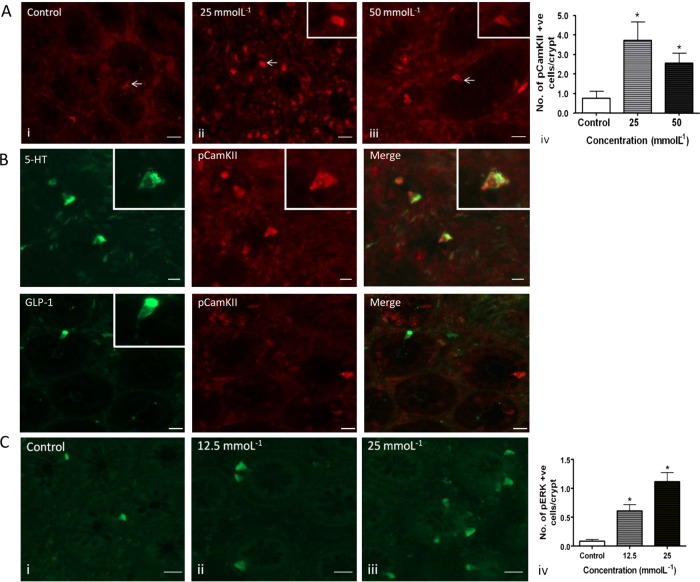
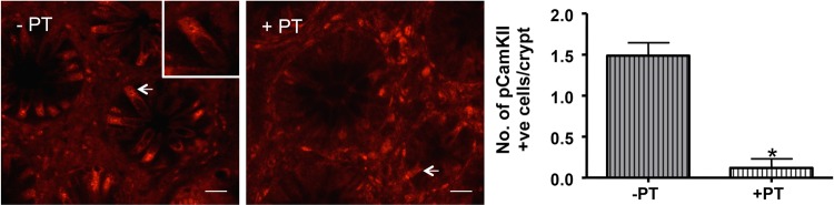
mRNA expression of 17 nutrient receptors and EEC mediators was assessed by quantitative PCR and found throughout mouse and human gut epithelium. Many species similarities emerged, in particular the dense expression of several receptors in the distal gut. Immunolabelling showed specific colocalisation of receptors with EEC mediators PYY and GLP-1 (L-cells) or 5-HT (enterochromaffin cells). We exposed isolated proximal colonic mucosa to specific nutrients, which recruited signalling pathways within specific EEC extracellular receptor-regulated kinase (p-ERK) and calmodulin kinase II (pCAMKII), as shown by subsequent immunolabelling, and activated release of these mediators. Aromatic amino acids activated both pathways in mouse, but in humans they induced only pCAMKII, which was colocalised mainly with 5-HT expression. Activation was pertussis toxin-sensitive. Fatty acid (C12) potently activated p-ERK in human in all EEC types and evoked potent release of all three mediators.
Specific nutrient receptors associate with distinct activation pathways within EEC. These may provide discrete, complementary pharmacological targets for intervention in obesity and type II diabetes.
当营养素刺激肠内分泌细胞(EEC)释放肠道激素时,食物摄入的抑制和葡萄糖稳态都会得到促进。几种特定的营养素受体可能位于EEC上,它们对膳食糖、氨基酸和脂肪酸作出反应。肥胖症和II型糖尿病的旁路手术通过将营养物质分流到肠道远端来发挥作用,在那里它会增加营养受体的激活和介质释放,但激活的细胞机制在很大程度上尚不清楚。我们确定了在小鼠和人类的哪些肠道区域以及哪些细胞中表达了哪些营养受体,它们如何与不同类型的EEC相关联,它们如何被激活从而导致激素和5-羟色胺(5-HT)释放。
通过定量PCR评估了17种营养受体和EEC介质的mRNA表达,并在小鼠和人类肠道上皮中均有发现。出现了许多物种间的相似性,特别是几种受体在肠道远端的密集表达。免疫标记显示受体与EEC介质酪酪肽(PYY)和胰高血糖素样肽-1(GLP-1)(L细胞)或5-HT(肠嗜铬细胞)有特异性共定位。我们将分离的近端结肠黏膜暴露于特定营养素,随后的免疫标记显示,这些营养素在特定的EEC细胞内募集了细胞外受体调节激酶(p-ERK)和钙调蛋白激酶II(pCAMKII)信号通路,并激活了这些介质的释放。芳香族氨基酸在小鼠中激活了这两条通路,但在人类中它们仅诱导pCAMKII,其主要与5-HT表达共定位。激活对百日咳毒素敏感。脂肪酸(C12)在所有EEC类型中均能有效激活人类的p-ERK,并引起所有三种介质的有效释放。
特定的营养受体与EEC内不同的激活途径相关联。这些可能为肥胖症和II型糖尿病的干预提供离散的、互补的药理学靶点。