• 文献检索
  • 文档翻译
  • 深度研究
  • 学术资讯
  • Suppr Zotero 插件Zotero 插件
  • 邀请有礼
  • 套餐&价格
  • 历史记录
应用&插件
Suppr Zotero 插件Zotero 插件浏览器插件Mac 客户端Windows 客户端微信小程序
定价
高级版会员购买积分包购买API积分包
服务
文献检索文档翻译深度研究API 文档MCP 服务
关于我们
关于 Suppr公司介绍联系我们用户协议隐私条款
关注我们

Suppr 超能文献

核心技术专利:CN118964589B侵权必究
粤ICP备2023148730 号-1Suppr @ 2026

文献检索

告别复杂PubMed语法,用中文像聊天一样搜索,搜遍4000万医学文献。AI智能推荐,让科研检索更轻松。

立即免费搜索

文件翻译

保留排版,准确专业,支持PDF/Word/PPT等文件格式,支持 12+语言互译。

免费翻译文档

深度研究

AI帮你快速写综述,25分钟生成高质量综述,智能提取关键信息,辅助科研写作。

立即免费体验

微栓子形成在兔急性蛛网膜下腔出血模型中的作用。

The role of microclot formation in an acute subarachnoid hemorrhage model in the rabbit.

机构信息

Department of Neurosurgery, Bern University Hospital, Inselspital Bern, 3012 Bern, Switzerland ; Laboratories for Neuroscience Research in Neurosurgery, Boston Children's Hospital, Boston, MA 02115, USA ; Harvard Medical School, Boston, MA 02115, USA.

Department of Intensive Care Medicine, Bern University Hospital, Inselspital Bern, 3012 Bern, Switzerland ; Department of Neurosurgery, University Hospital Cologne, 50924 Cologne, Germany.

出版信息

Biomed Res Int. 2014;2014:161702. doi: 10.1155/2014/161702. Epub 2014 Jul 7.

DOI:10.1155/2014/161702
PMID:25110658
原文链接:https://pmc.ncbi.nlm.nih.gov/articles/PMC4109416/
Abstract

BACKGROUND

Microvascular dysfunction and microthrombi formation are believed to contribute to development of early brain injury (EBI) after aneurysmal subarachnoid hemorrhage (SAH).

OBJECTIVE

This study aimed to determine (i) extent of microthrombus formation and neuronal apoptosis in the brain parenchyma using a blood shunt SAH model in rabbits; (ii) correlation of structural changes in microvessels with EBI characteristics.

METHODS

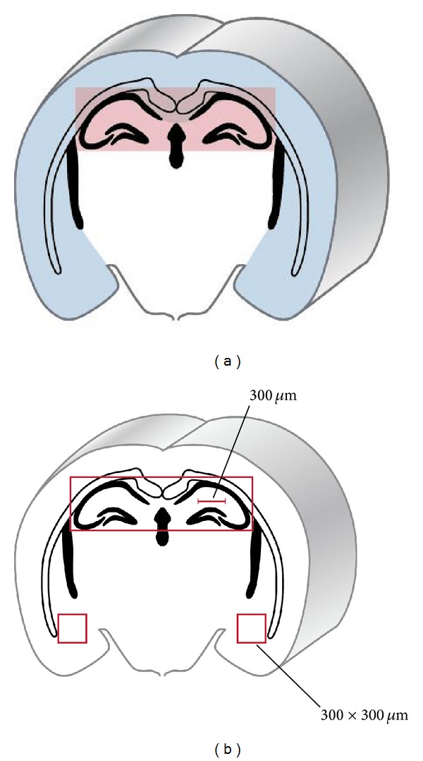
Acute SAH was induced using a rabbit shunt cisterna magna model. Extent of microthrombosis was detected 24 h post-SAH (n = 8) by fibrinogen immunostaining, compared to controls (n = 4). We assessed apoptosis by terminal deoxynucleotidyl transferase nick end labeling (TUNEL) in cortex and hippocampus.

RESULTS

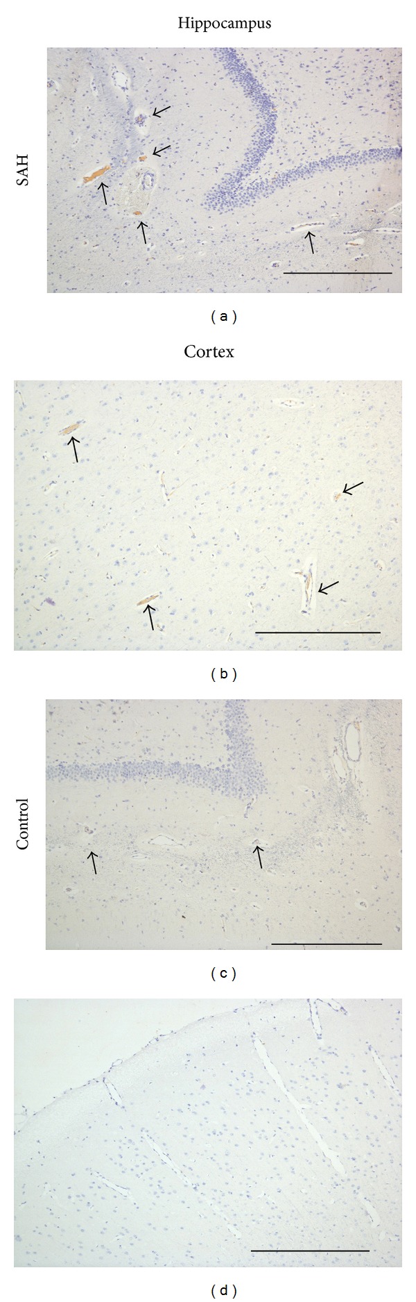
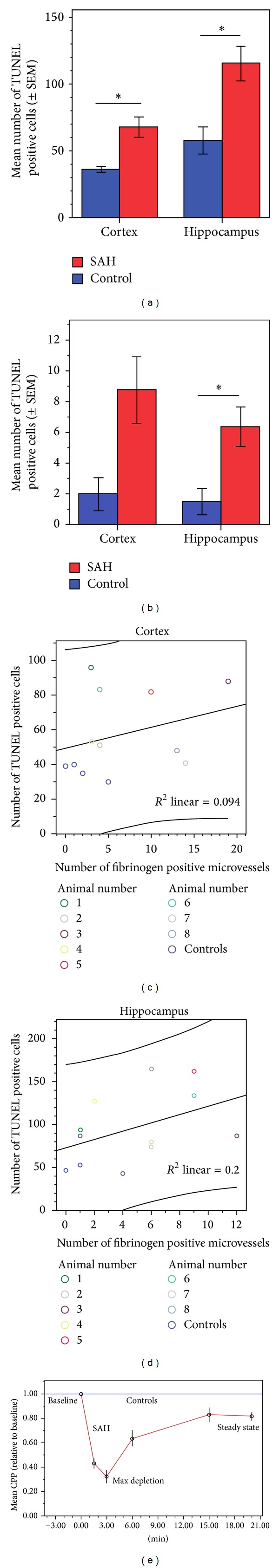
Our results showed significantly more TUNEL-positive cells (SAH: 115 ± 13; controls: 58 ± 10; P = 0.016) and fibrinogen-positive microthromboemboli (SAH: 9 ± 2; controls: 2 ± 1; P = 0.03) in the hippocampus after aneurysmal SAH.

CONCLUSIONS

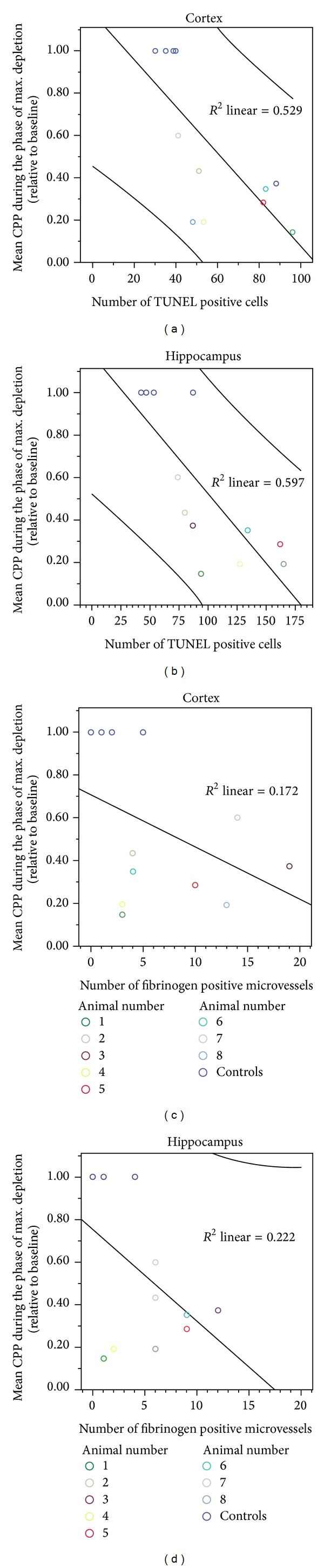
We found clear evidence of early microclot formation in a rabbit model of acute SAH. The extent of microthrombosis did not correlate with early apoptosis or CPP depletion after SAH; however, the total number of TUNEL positive cells in the cortex and the hippocampus significantly correlated with mean CPP reduction during the phase of maximum depletion after SAH induction. Both microthrombosis and neuronal apoptosis may contribute to EBI and subsequent DCI.

摘要

背景

微血管功能障碍和微血栓形成被认为是导致蛛网膜下腔出血(SAH)后早期脑损伤(EBI)的原因。

目的

本研究旨在:(i)使用兔血分流蛛网膜下腔出血模型确定脑实质中微血栓形成和神经元凋亡的程度;(ii)研究微血管结构变化与 EBI 特征的相关性。

方法

采用兔分流枕大池模型诱导急性 SAH。通过纤维蛋白原免疫染色检测 SAH 后 24 小时(n = 8)与对照组(n = 4)相比微血栓形成的程度。通过末端脱氧核苷酸转移酶缺口末端标记法(TUNEL)评估皮质和海马中的细胞凋亡。

结果

结果显示,SAH 组海马区 TUNEL 阳性细胞(SAH:115 ± 13;对照组:58 ± 10;P = 0.016)和纤维蛋白原阳性微血栓(SAH:9 ± 2;对照组:2 ± 1;P = 0.03)明显多于对照组。

结论

我们在兔急性 SAH 模型中发现了早期微栓子形成的明确证据。微血栓形成的程度与 SAH 后早期细胞凋亡或 CPP 耗竭无关;然而,皮质和海马中 TUNEL 阳性细胞的总数与 SAH 诱导后 CPP 最大耗竭期间的平均 CPP 降低显著相关。微血栓形成和神经元凋亡可能导致 EBI 和随后的 DCI。

https://cdn.ncbi.nlm.nih.gov/pmc/blobs/436c/4109416/d598590a6f75/BMRI2014-161702.004.jpg
https://cdn.ncbi.nlm.nih.gov/pmc/blobs/436c/4109416/bf0cd0ecc922/BMRI2014-161702.001.jpg
https://cdn.ncbi.nlm.nih.gov/pmc/blobs/436c/4109416/07719c7b8719/BMRI2014-161702.002.jpg
https://cdn.ncbi.nlm.nih.gov/pmc/blobs/436c/4109416/bdae582cc0f0/BMRI2014-161702.003.jpg
https://cdn.ncbi.nlm.nih.gov/pmc/blobs/436c/4109416/d598590a6f75/BMRI2014-161702.004.jpg

相似文献

1
The role of microclot formation in an acute subarachnoid hemorrhage model in the rabbit.微栓子形成在兔急性蛛网膜下腔出血模型中的作用。
Biomed Res Int. 2014;2014:161702. doi: 10.1155/2014/161702. Epub 2014 Jul 7.
2
A new rabbit model for the study of early brain injury after subarachnoid hemorrhage.蛛网膜下腔出血后早期脑损伤的新型兔模型。
J Neurosci Methods. 2012 Jul 15;208(2):138-45. doi: 10.1016/j.jneumeth.2012.05.010. Epub 2012 May 14.
3
Tocilizumab Reduces Vasospasms, Neuronal Cell Death, and Microclot Formation in a Rabbit Model of Subarachnoid Hemorrhage.托珠单抗可减少蛛网膜下腔出血兔模型的血管痉挛、神经元细胞死亡和微栓形成。
Transl Stroke Res. 2021 Oct;12(5):894-904. doi: 10.1007/s12975-020-00880-3. Epub 2021 Jan 6.
4
Neuroprotective effect of hydrogen-rich saline against neurologic damage and apoptosis in early brain injury following subarachnoid hemorrhage: possible role of the Akt/GSK3β signaling pathway.富氢生理盐水对蛛网膜下腔出血后早期脑损伤神经损伤和细胞凋亡的神经保护作用:可能与 Akt/GSK3β信号通路有关。
PLoS One. 2014 Apr 24;9(4):e96212. doi: 10.1371/journal.pone.0096212. eCollection 2014.
5
Changes in the cerebrospinal fluid lipid profile following subarachnoid hemorrhage in a closed cranium model: Correlations to cerebral vasospasm, neuronal cell death and Interleukin-6 synthesis. A pilot study.闭合颅骨模型下蛛网膜下腔出血后脑脊液脂质谱的变化:与脑血管痉挛、神经元细胞死亡及白细胞介素-6合成的相关性。一项初步研究。
J Stroke Cerebrovasc Dis. 2020 Sep;29(9):105054. doi: 10.1016/j.jstrokecerebrovasdis.2020.105054. Epub 2020 Jun 23.
6
Mechanisms of microthrombi formation after experimental subarachnoid hemorrhage.实验性蛛网膜下腔出血后微血栓形成的机制。
Neuroscience. 2012 Nov 8;224:26-37. doi: 10.1016/j.neuroscience.2012.08.002. Epub 2012 Aug 16.
7
Mechanisms of microthrombosis and microcirculatory constriction after experimental subarachnoid hemorrhage.实验性蛛网膜下腔出血后微血栓形成及微循环收缩的机制
Acta Neurochir Suppl. 2013;115:185-92. doi: 10.1007/978-3-7091-1192-5_35.
8
[Cerebral lymphatic blockage aggravates apoptosis of cortical neurons after subarachnoid hemorrhage in rats in vivo].[脑淋巴引流阻滞加重大鼠蛛网膜下腔出血后皮质神经元的体内凋亡]
Sheng Li Xue Bao. 2010 Apr 25;62(2):109-14.
9
Deficiency of Tenascin-C Alleviates Neuronal Apoptosis and Neuroinflammation After Experimental Subarachnoid Hemorrhage in Mice.Tenascin-C 缺乏可减轻实验性蛛网膜下腔出血后小鼠的神经元凋亡和神经炎症。
Mol Neurobiol. 2018 Nov;55(11):8346-8354. doi: 10.1007/s12035-018-1006-z. Epub 2018 Mar 15.
10
Ethyl Pyruvate Attenuates Early Brain Injury Following Subarachnoid Hemorrhage in the Endovascular Perforation Rabbit Model Possibly Via Anti-inflammation and Inhibition of JNK Signaling Pathway.丙酮酸乙酯可能通过抗炎和抑制JNK信号通路减轻血管内穿刺兔模型蛛网膜下腔出血后的早期脑损伤。
Neurochem Res. 2017 Apr;42(4):1044-1056. doi: 10.1007/s11064-016-2138-z. Epub 2017 Feb 25.

引用本文的文献

1
Role of microcirculatory impairment in delayed cerebral ischemia and outcome after aneurysmal subarachnoid hemorrhage.微血管损伤在颅内动脉瘤性蛛网膜下腔出血后迟发性脑缺血及转归中的作用。
J Cereb Blood Flow Metab. 2022 Jan;42(1):186-196. doi: 10.1177/0271678X211045446. Epub 2021 Sep 9.
2
Preclinical and clinical role of interleukin-6 in the development of delayed cerebral vasospasm and neuronal cell death after subarachnoid hemorrhage: towards a potential target therapy?白介素-6 在蛛网膜下腔出血后迟发性脑血管痉挛和神经元细胞死亡发展中的临床前和临床作用:潜在的靶向治疗?
Neurosurg Rev. 2022 Feb;45(1):395-403. doi: 10.1007/s10143-021-01628-9. Epub 2021 Aug 27.
3

本文引用的文献

1
Effect of ADAMTS-13 on cerebrovascular microthrombosis and neuronal injury after experimental subarachnoid hemorrhage.ADAMTS-13 对实验性蛛网膜下腔出血后脑血管微血栓形成和神经元损伤的影响。
J Thromb Haemost. 2014 Apr;12(4):505-14. doi: 10.1111/jth.12511.
2
Delayed neurological deterioration after subarachnoid haemorrhage.蛛网膜下腔出血后神经功能恶化延迟。
Nat Rev Neurol. 2014 Jan;10(1):44-58. doi: 10.1038/nrneurol.2013.246. Epub 2013 Dec 10.
3
FMRI of working memory impairment after recovery from subarachnoid hemorrhage.蛛网膜下腔出血后恢复期间工作记忆损伤的 fMRI 研究。
Role of platelets in the pathogenesis of delayed injury after subarachnoid hemorrhage.
血小板在蛛网膜下腔出血后迟发性损伤发病机制中的作用。
J Cereb Blood Flow Metab. 2021 Nov;41(11):2820-2830. doi: 10.1177/0271678X211020865. Epub 2021 Jun 10.
4
Neuroprotective Strategies in Aneurysmal Subarachnoid Hemorrhage (aSAH).动脉瘤性蛛网膜下腔出血(aSAH)的神经保护策略。
Int J Mol Sci. 2021 May 21;22(11):5442. doi: 10.3390/ijms22115442.
5
Microvascular platelet aggregation and thrombosis after subarachnoid hemorrhage: A review and synthesis.蛛网膜下腔出血后微血管血小板聚集和血栓形成:综述与综合。
J Cereb Blood Flow Metab. 2020 Aug;40(8):1565-1575. doi: 10.1177/0271678X20921974. Epub 2020 Apr 28.
6
The Role of Sartans in the Treatment of Stroke and Subarachnoid Hemorrhage: A Narrative Review of Preclinical and Clinical Studies.沙坦类药物在中风和蛛网膜下腔出血治疗中的作用:临床前和临床研究的叙述性综述
Brain Sci. 2020 Mar 7;10(3):153. doi: 10.3390/brainsci10030153.
7
The Role of Thromboinflammation in Delayed Cerebral Ischemia after Subarachnoid Hemorrhage.血栓炎症在蛛网膜下腔出血后迟发性脑缺血中的作用
Front Neurol. 2017 Oct 23;8:555. doi: 10.3389/fneur.2017.00555. eCollection 2017.
8
Original Research: Establishment of an early embolus-related cerebral injury model after cardiopulmonary bypass in miniature pigs.原创研究:小型猪体外循环后早期栓子相关性脑损伤模型的建立
Exp Biol Med (Maywood). 2016 Oct;241(16):1819-24. doi: 10.1177/1535370216648804. Epub 2016 May 5.
9
Early brain injury linearly correlates with reduction in cerebral perfusion pressure during the hyperacute phase of subarachnoid hemorrhage.在蛛网膜下腔出血超急性期,早期脑损伤与脑灌注压降低呈线性相关。
Intensive Care Med Exp. 2014 Dec;2(1):30. doi: 10.1186/s40635-014-0030-1. Epub 2014 Nov 30.
10
The rabbit shunt model of subarachnoid haemorrhage.兔蛛网膜下腔出血分流模型。
Transl Stroke Res. 2014 Dec;5(6):669-80. doi: 10.1007/s12975-014-0369-3. Epub 2014 Oct 19.
Front Neurol. 2013 Nov 6;4:179. doi: 10.3389/fneur.2013.00179. eCollection 2013.
4
Controversies and evolving new mechanisms in subarachnoid hemorrhage.蛛网膜下腔出血的争议与新机制的演变
Prog Neurobiol. 2014 Apr;115:64-91. doi: 10.1016/j.pneurobio.2013.09.002. Epub 2013 Sep 25.
5
Neutrophils express oncomodulin and promote optic nerve regeneration.中性粒细胞表达骨桥蛋白并促进视神经再生。
J Neurosci. 2013 Sep 11;33(37):14816-24. doi: 10.1523/JNEUROSCI.5511-12.2013.
6
Early cerebral infarction following aneurysmal subarachnoid hemorrhage: frequency, risk factors, patterns, and prognosis.颅内破裂动脉瘤性蛛网膜下腔出血后早期脑梗死:频率、危险因素、类型和预后。
Curr Neurovasc Res. 2013 Nov;10(4):316-24. doi: 10.2174/15672026113109990027.
7
Nasal administration of recombinant osteopontin attenuates early brain injury after subarachnoid hemorrhage.鼻腔给予重组骨桥蛋白可减轻蛛网膜下腔出血后的早期脑损伤。
Stroke. 2013 Nov;44(11):3189-94. doi: 10.1161/STROKEAHA.113.001574. Epub 2013 Sep 5.
8
Early brain injury, an evolving frontier in subarachnoid hemorrhage research.早期脑损伤:蛛网膜下腔出血研究的不断发展前沿。
Transl Stroke Res. 2013 Aug;4(4):432-46. doi: 10.1007/s12975-013-0257-2.
9
Impact of global cerebral atrophy on clinical outcome after subarachnoid hemorrhage.全球脑萎缩对蛛网膜下腔出血后临床结局的影响。
J Neurosurg. 2013 Jul;119(1):198-206. doi: 10.3171/2013.3.JNS121950. Epub 2013 May 10.
10
Granulocyte-colony stimulating factor in combination with stem cell factor confers greater neuroprotection after hypoxic-ischemic brain damage in the neonatal rats than a solitary treatment.粒细胞集落刺激因子与干细胞因子联合使用对新生大鼠缺氧缺血性脑损伤后的神经保护作用比单独使用更强。
Transl Stroke Res. 2013 Apr;4(2):171-8. doi: 10.1007/s12975-012-0225-2. Epub 2012 Nov 8.