Lentz Margaret R, Peterson Kristin L, Ibrahim Wael G, Lee Dianne E, Sarlls Joelle, Lizak Martin J, Maric Dragan, Reid William C, Hammoud Dima A
Center for Infectious Disease Imaging (CIDI), Radiology and Imaging Sciences, Clinical Center, National Institutes of Health (NIH), Bethesda, Maryland, United States of America.
National Institute of Child Health and Human Development (NICHD), NIH, Bethesda, Maryland, United States of America.
PLoS One. 2014 Aug 21;9(8):e105752. doi: 10.1371/journal.pone.0105752. eCollection 2014.
There are currently no widely accepted neuro-HIV small animal models. We wanted to validate the HIV-1 Transgenic rat (Tg) as an appropriate neuro-HIV model and then establish in vivo imaging biomarkers of neuropathology, within this model, using MR structural and diffusion tensor imaging (DTI).
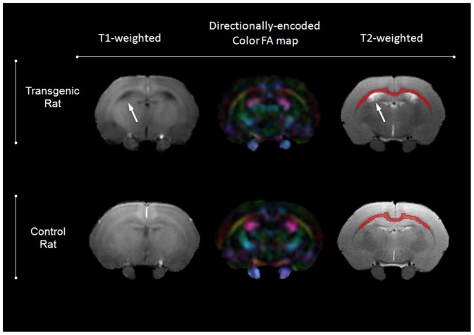
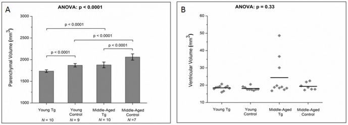
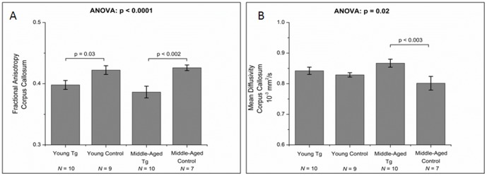
Young and middle-aged Tg and control rats were imaged using MRI. A subset of middle-aged animals underwent longitudinal repeat imaging six months later. Total brain volume (TBV), ventricular volume (VV) and parenchymal volume (PV = TBV-VV) were measured. Fractional anisotropy (FA) and mean diffusivity (MD) values of the corpus callosum (CC) were calculated from DTI data.
TBV and PV were smaller in Tg compared to control rats in young and middle-aged cohorts (p<0.0001). VV increased significantly (p = 0.005) over time in the longitudinal Tg cohort. There were lower FA (p<0.002) and higher MD (p<0.003) values in the CC of middle-aged Tg rats compared to age-matched controls. Longitudinally, MD significantly decreased over time in Tg rats (p<0.03) while it did not change significantly in the control cohort over the same period of time (p>0.05).
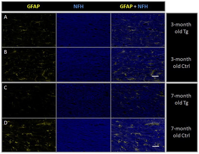
We detected brain volume loss in the Tg rat, probably due to astrocytic dysfunction/loss, loss of structural/axonal matrix and striatal neuronal loss as suggested by immunofluorescence. Increased MD and decreased FA in the CC probably reflect microstructural differences between the Tg and Control rats which could include increased extracellular space between white matter tracts, demyelination and axonal degeneration, among other pathologies. We believe that the Tg rat is an adequate model of neuropathology in HIV and that volumetric MR and DTI measures can be potentially used as biomarkers of disease progression.
目前尚无被广泛接受的神经艾滋病小动物模型。我们希望验证HIV-1转基因大鼠(Tg)作为合适的神经艾滋病模型,然后在该模型中使用磁共振结构成像和扩散张量成像(DTI)建立神经病理学的体内成像生物标志物。
对年轻和中年Tg大鼠及对照大鼠进行磁共振成像。一部分中年动物在6个月后接受纵向重复成像。测量全脑体积(TBV)、脑室体积(VV)和实质体积(PV = TBV - VV)。从DTI数据计算胼胝体(CC)的各向异性分数(FA)和平均扩散率(MD)值。
在年轻和中年队列中,Tg大鼠的TBV和PV均小于对照大鼠(p<0.0001)。纵向Tg队列中,VV随时间显著增加(p = 0.005)。与年龄匹配的对照相比,中年Tg大鼠CC中的FA值较低(p<0.002),MD值较高(p<0.003)。纵向来看,Tg大鼠的MD随时间显著降低(p<0.03),而同期对照队列中MD无显著变化(p>0.05)。
我们在Tg大鼠中检测到脑体积减少,可能是由于免疫荧光显示的星形细胞功能障碍/丧失、结构/轴突基质丧失和纹状体神经元丧失。CC中MD增加和FA降低可能反映了Tg大鼠与对照大鼠之间的微观结构差异,这可能包括白质束之间细胞外间隙增加、脱髓鞘和轴突变性等其他病理情况。我们认为Tg大鼠是艾滋病神经病理学的合适模型,容积磁共振成像和DTI测量有可能用作疾病进展的生物标志物。