Stoffels Josephine M J, Hoekstra Dick, Franklin Robin J M, Baron Wia, Zhao Chao
Department of Cell Biology, University of Groningen, University Medical Center Groningen, The Netherlands.
Glia. 2015 Feb;63(2):242-56. doi: 10.1002/glia.22748. Epub 2014 Aug 25.
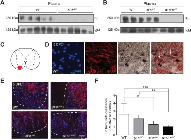
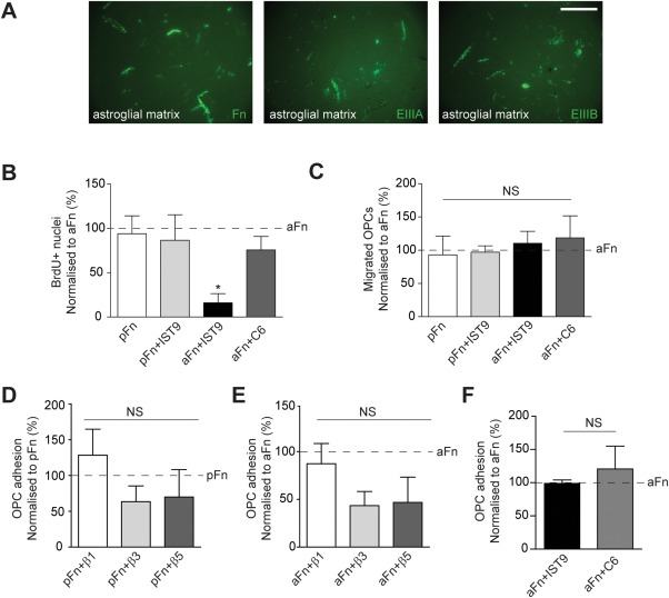
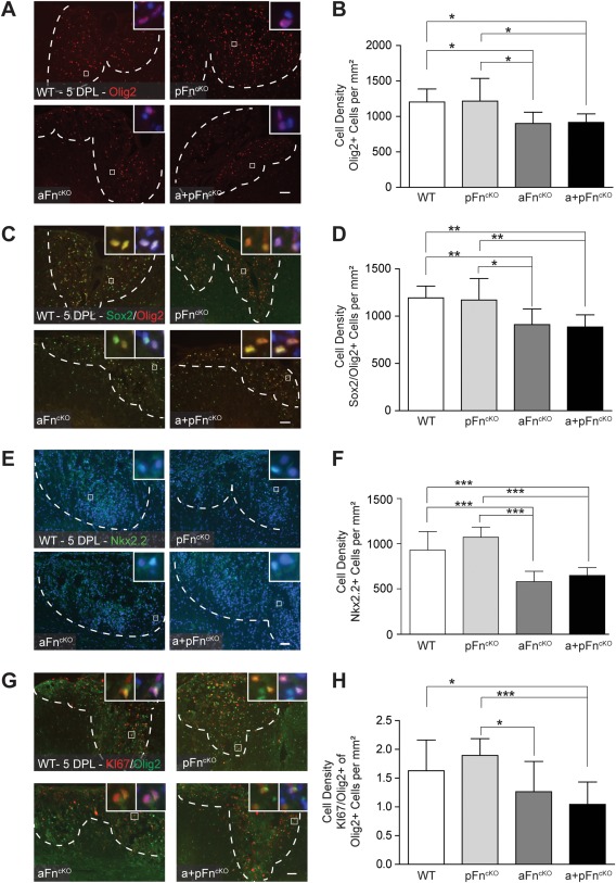
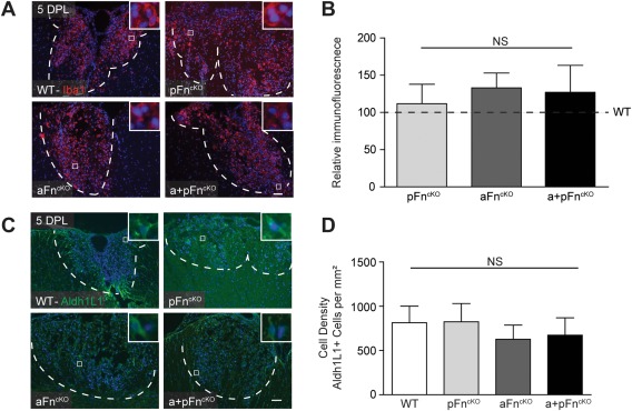
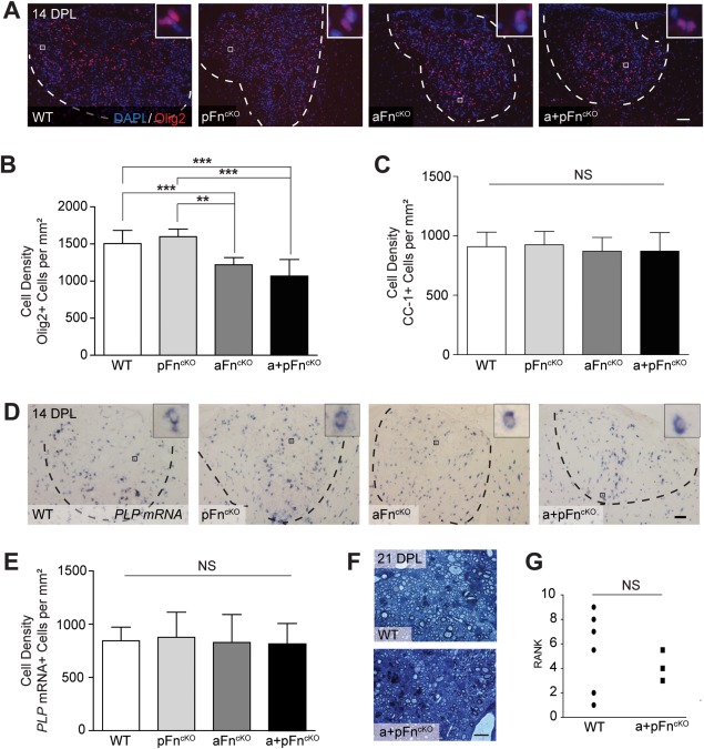
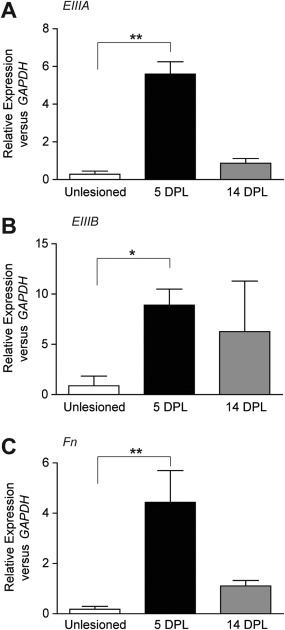
Central nervous system remyelination by oligodendrocyte progenitor cells (OPCs) ultimately fails in the majority of multiple sclerosis (MS) lesions. Remyelination benefits from transient expression of factors that promote migration and proliferation of OPCs, which may include fibronectin (Fn). Fn is present in demyelinated lesions in two major forms; plasma Fn (pFn), deposited following blood-brain barrier disruption, and cellular Fn, synthesized by resident glial cells and containing alternatively spliced domains EIIIA and EIIIB. Here, we investigated the distinctive roles that astrocyte-derived Fn (aFn) and pFn play in remyelination. We used an inducible Cre-lox recombination strategy to selectively remove pFn, aFn or both from mice, and examined the impact on remyelination of toxin-induced demyelinated lesions of spinal cord white matter. This approach revealed that astrocytes are a major source of Fn in demyelinated lesions. Furthermore, following aFn conditional knockout, the number of OPCs recruited to the demyelinated lesion decreased significantly, whereas OPC numbers were unaltered following pFn conditional knockout. However, remyelination completed normally following conditional knockout of aFn and pFn. Both the EIIIA and EIIIB domains of aFn were expressed following demyelination, and in vitro assays demonstrated that the EIIIA domain of aFn mediates proliferation of OPCs, but not migration. Therefore, although the EIIIA domain from aFn mediates OPC proliferation, aFn is not essential for successful remyelination. Since previous findings indicated that astrocyte-derived Fn aggregates in chronic MS lesions inhibit remyelination, aFn removal may benefit therapeutic strategies to promote remyelination in MS.
少突胶质前体细胞(OPCs)对中枢神经系统的重新髓鞘化在大多数多发性硬化症(MS)病变中最终会失败。重新髓鞘化受益于促进OPCs迁移和增殖的因子的短暂表达,其中可能包括纤连蛋白(Fn)。Fn以两种主要形式存在于脱髓鞘病变中;血浆Fn(pFn),在血脑屏障破坏后沉积,以及细胞Fn,由驻留神经胶质细胞合成并包含选择性剪接的结构域EIIIA和EIIIB。在这里,我们研究了星形胶质细胞衍生的Fn(aFn)和pFn在重新髓鞘化中所起的独特作用。我们使用诱导性Cre-lox重组策略从小鼠中选择性去除pFn、aFn或两者,并检查对脊髓白质毒素诱导的脱髓鞘病变重新髓鞘化的影响。这种方法表明星形胶质细胞是脱髓鞘病变中Fn的主要来源。此外,在aFn条件性敲除后,招募到脱髓鞘病变中的OPCs数量显著减少,而在pFn条件性敲除后OPCs数量未改变。然而,在aFn和pFn条件性敲除后重新髓鞘化正常完成。脱髓鞘后aFn的EIIIA和EIIIB结构域均有表达,体外实验表明aFn的EIIIA结构域介导OPCs的增殖,但不介导迁移。因此,尽管aFn的EIIIA结构域介导OPCs增殖,但aFn对于成功的重新髓鞘化并非必不可少。由于先前的研究结果表明慢性MS病变中星形胶质细胞衍生的Fn聚集体会抑制重新髓鞘化,去除aFn可能有利于促进MS重新髓鞘化的治疗策略。