Handgraaf Henricus J M, Boonstra Martin C, Van Erkel Arian R, Bonsing Bert A, Putter Hein, Van De Velde Cornelis J H, Vahrmeijer Alexander L, Mieog J Sven D
Department of Surgery, Leiden University Medical Center, Albinusdreef 2, 2300 RC Leiden, The Netherlands.
Department of Radiology, Leiden University Medical Center, Albinusdreef 2, 2300 RC Leiden, The Netherlands.
Biomed Res Int. 2014;2014:890230. doi: 10.1155/2014/890230. Epub 2014 Jul 15.
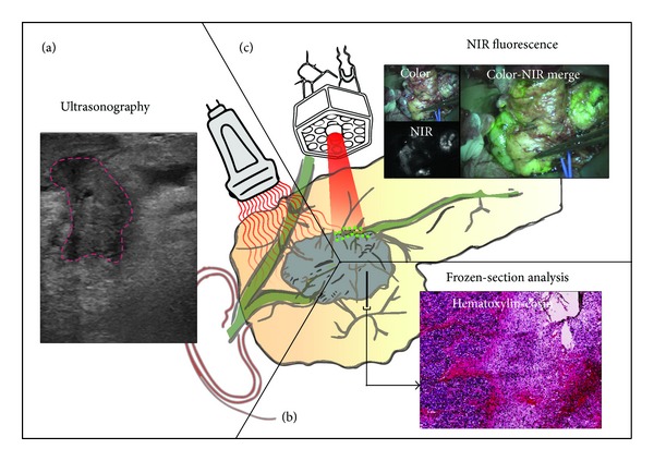
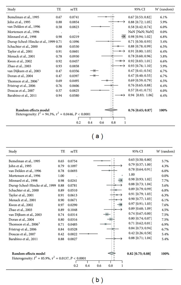
Prognosis of patients with pancreatic cancer is poor. Even the small minority that undergoes resection with curative intent has low 5-year survival rates. This may partly be explained by the high number of irradical resections, which results in local recurrence and impaired overall survival. Currently, ultrasonography is used during surgery for resectability assessment and frozen-section analysis is used for assessment of resection margins in order to decrease the number of irradical resections. The introduction of minimal invasive techniques in pancreatic surgery has deprived surgeons from direct tactile information. To improve intraoperative assessment of pancreatic tumor extension, enhanced or novel intraoperative imaging technologies accurately visualizing and delineating cancer cells are necessary. Emerging modalities are intraoperative near-infrared fluorescence imaging and freehand nuclear imaging using tumor-specific targeted contrast agents. In this review, we performed a meta-analysis of the literature on laparoscopic ultrasonography and we summarized and discussed current and future intraoperative imaging modalities and their potential for improved tumor demarcation during pancreatic surgery.
胰腺癌患者的预后较差。即使是少数接受根治性切除的患者,其5年生存率也很低。这部分可能是由于大量的非根治性切除,导致局部复发并损害总生存率。目前,手术期间使用超声检查进行可切除性评估,使用冰冻切片分析评估切缘,以减少非根治性切除的数量。胰腺手术中微创技术的引入使外科医生失去了直接的触觉信息。为了改善术中对胰腺肿瘤扩展的评估,需要增强或新颖的术中成像技术来准确可视化和描绘癌细胞。新兴的模式是术中近红外荧光成像和使用肿瘤特异性靶向造影剂的徒手核成像。在本综述中,我们对腹腔镜超声检查的文献进行了荟萃分析,并总结和讨论了当前和未来的术中成像模式及其在胰腺手术中改善肿瘤边界划分的潜力。