Galic Jerman Katarina, Kobal Borut, Jakimovska Marina, Verdenik Ivan, Cerne Katarina
Institute of Pharmacology and Experimental Toxicology, Faculty of Medicine, University Ljubljana, Korytkova 2, 1000 Ljubljana, Slovenia.
World J Surg Oncol. 2014 Sep 4;12:278. doi: 10.1186/1477-7819-12-278.
Determination of the tumor marker concentration in peritoneal fluid (PF) may help to assess its potential to detect small concentration changes between benign ovarian pathology and early stage ovarian cancer. Peritoneal washing, which can also be obtained when PF is absent, is already included in the International Federation of Gynecology and Obstetrics (FIGO) staging classification for ovarian cancer but sampling has not yet been standardized. Since our aim was to evaluate the relationship between marker concentration in PF and washing, standardization of the sampling protocol was a prerequisite to ensure reliable results.
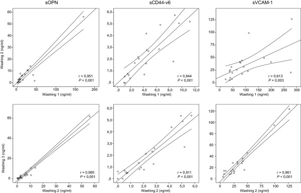
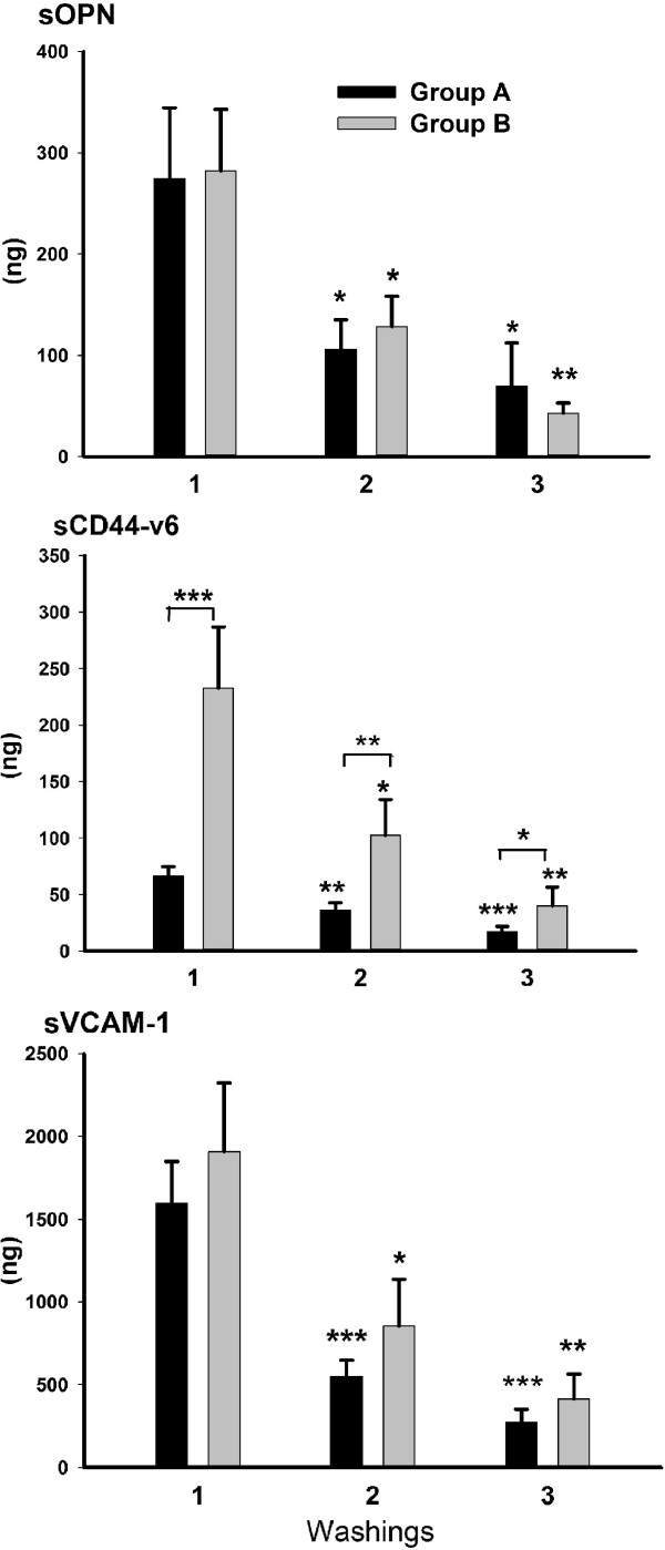
Thirty-three women with non-malignant pathology of the reproductive organs were included in the study. We used three promising tumor markers for evaluation of the marker concentration in local fluid: osteopontin (sOPN), splice variant 6 of sCD44 (sCD44-v6) and vascular cell adhesion molecule-1 (sVCAM-1). After aspiration of PF, washing of the uterus, ovaries and pelvic peritoneum was performed with saline solution. Patients were divided into two groups based on the solution volume: A-20 ml and B-50 ml. To determine the efficiency of washing in relation to solution volume, washing was repeated three times. Concentrations of markers in samples were determined using flow cytometry.
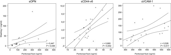
Mean concentrations of markers were significantly higher (P <0.001) in PF than in the first washing. We demonstrated a significant positive correlation between marker concentrations in PF and first washing (sOPN: r = 0.447, P = 0.048; sCD44-v6: r = 0.660, P = 0.002; sVCAM-1: r = 0.526, P = 0.017). When using a smaller solution volume for washing, significantly higher (sVCAM-1: 2.5-fold, P = 0.021; sOPN: 3-fold, P = 0.024) or equal (sCD44-v6) mean concentrations of tumor markers were obtained.
Our work demonstrates for the first time that concentrations of sOPN, sCD44-v6 and sVCAM-1 in PF correlate with peritoneal washing in women with non-malignant pathology of the reproductive organs. This indicates that, for selected tumor markers, washing can replace PF when PF is absent. A standardized protocol for sampling PF and performing washing during laparoscopy was established.
测定腹腔液(PF)中的肿瘤标志物浓度可能有助于评估其检测良性卵巢病变与早期卵巢癌之间微小浓度变化的潜力。国际妇产科联合会(FIGO)的卵巢癌分期分类中已纳入腹腔冲洗(当没有PF时也可进行),但采样尚未标准化。由于我们的目的是评估PF和冲洗液中标志物浓度之间的关系,因此采样方案的标准化是确保可靠结果的先决条件。
33名患有生殖器官非恶性病变的女性被纳入研究。我们使用三种有前景的肿瘤标志物来评估局部液体中的标志物浓度:骨桥蛋白(sOPN)、sCD44的剪接变体6(sCD44-v6)和血管细胞黏附分子-1(sVCAM-1)。抽取PF后,用盐溶液冲洗子宫、卵巢和盆腔腹膜。根据溶液体积将患者分为两组:A组-20毫升和B组-50毫升。为了确定冲洗效率与溶液体积的关系,冲洗重复三次。使用流式细胞术测定样本中标志物的浓度。
PF中标志物的平均浓度显著高于首次冲洗液(P<0.001)。我们证明了PF和首次冲洗液中标志物浓度之间存在显著正相关(sOPN:r = 0.447,P = 0.048;sCD44-v6:r = 0.660,P = 0.002;sVCAM-1:r = 0.526,P = 0.017)。当使用较小体积的溶液进行冲洗时,肿瘤标志物的平均浓度显著更高(sVCAM-1:2.5倍,P = 0.021;sOPN:3倍,P = 0.024)或相等(sCD44-v6)。
我们的研究首次表明,患有生殖器官非恶性病变的女性PF中sOPN、sCD44-v6和sVCAM-1的浓度与腹腔冲洗液相关。这表明,对于选定的肿瘤标志物,如果没有PF,冲洗液可以替代PF。建立了腹腔镜检查期间采集PF和进行冲洗的标准化方案。