Binch Abbie L A, Cole Ashley A, Breakwell Lee M, Michael Anthony L R, Chiverton Neil, Cross Alison K, Le Maitre Christine L
Arthritis Res Ther. 2014 Aug 20;16(5):416. doi: 10.1186/s13075-014-0416-1.
The degenerate intervertebral disc (IVD) becomes innervated by sensory nerve fibres, and vascularised by blood vessels. This study aimed to identify neurotrophins, neuropeptides and angiogenic factors within native IVD tissue and to further investigate whether pro-inflammatory cytokines are involved in the regulation of expression levels within nucleus pulposus (NP) cells, nerve and endothelial cells.
Quantitative real-time PCR (qRT-PCR) was performed on 53 human IVDs from 52 individuals to investigate native gene expression of neurotrophic factors and their receptors, neuropeptides and angiogenic factors. The regulation of these factors by cytokines was investigated in NP cells in alginate culture, and nerve and endothelial cells in monolayer using RT-PCR and substance P (SP) protein expression in interleukin-1 (IL-1β) stimulated NP cells.
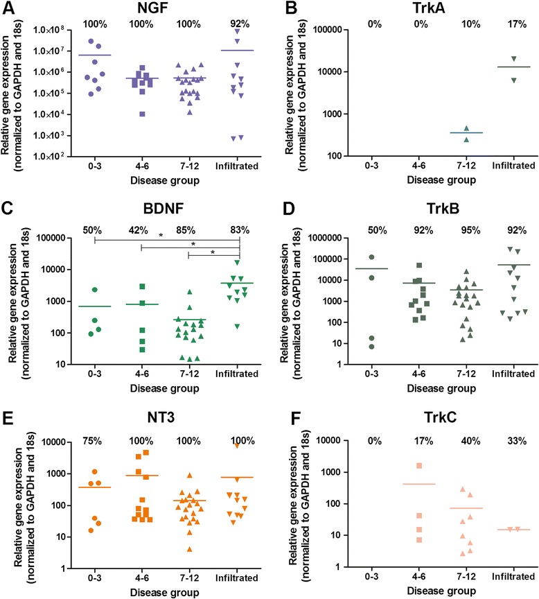
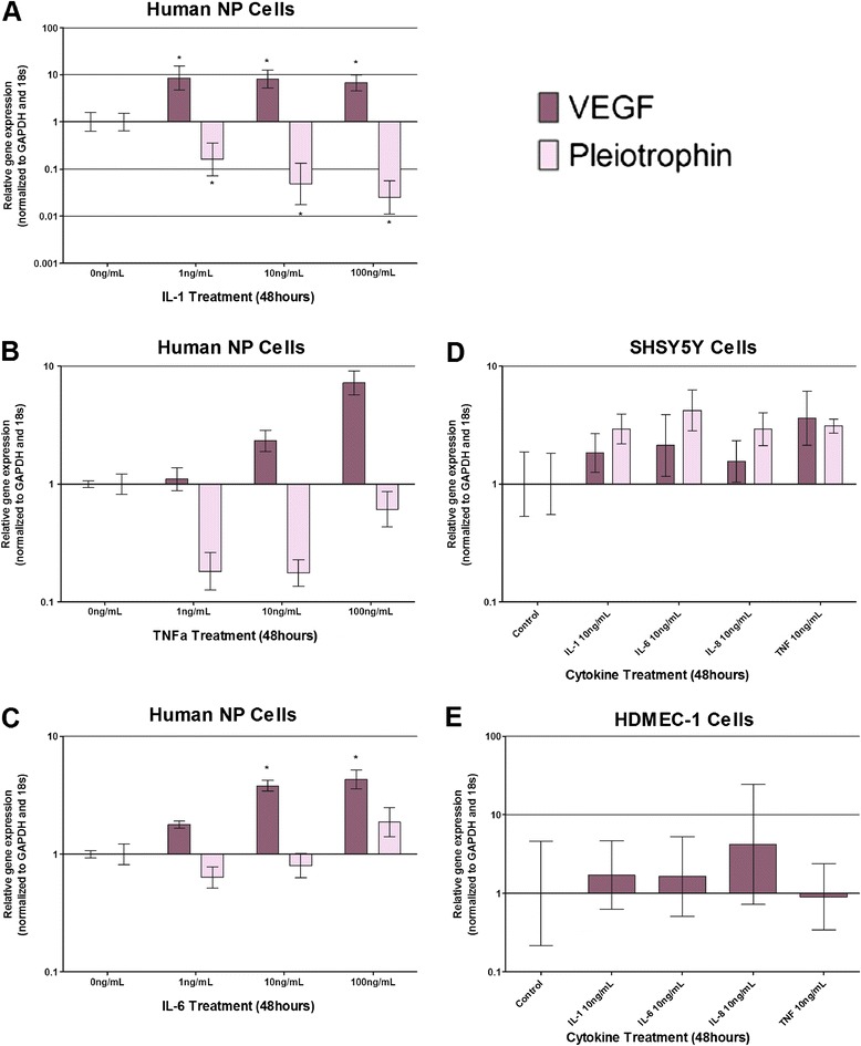
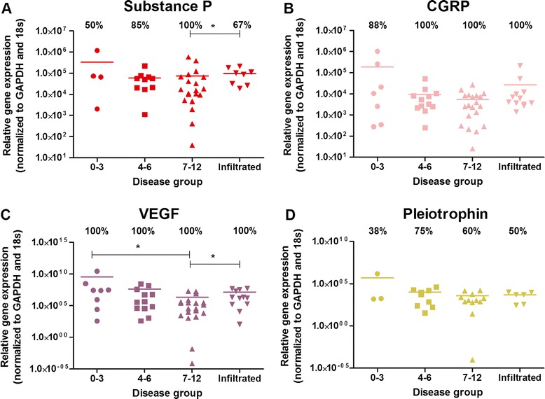
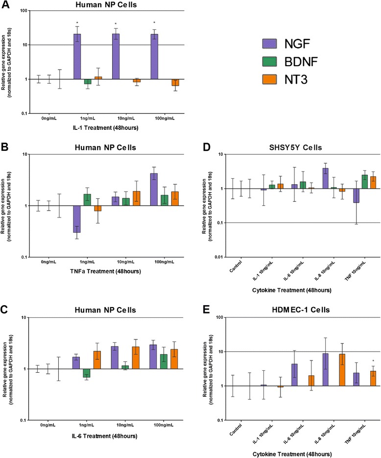
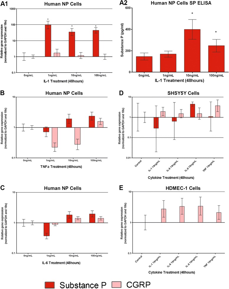
Initial investigation on uncultured NP cells identified expression of all neurotrophins by native NP cells, whilst the nerve growth factor (NGF) receptor was only identified in severely degenerate and infiltrated discs, and brain derived neurotrophic factor (BDNF) receptor expressed by more degenerate discs. BDNF expression was significantly increased in infiltrated and degenerate samples. SP and vascular endothelial growth factor (VEGF) were higher in infiltrated samples. In vitro stimulation by IL-1β induced NGF in NP cells. Neurotropin-3 was induced by tumour necrosis factor alpha in human dermal microvascular endothelial cells (HDMECs). SP gene and protein expression was increased in NP cells by IL-1β. Calcitonin gene related peptide was increased in SH-SY5Y cells upon cytokine stimulation. VEGF was induced by IL-1β and interleukin-6 in NP cells, whilst pleiotrophin was decreased by IL-1β. VEGF and pleiotrophin were expressed by SH-SY5Y cells, and VEGF by HDMECs, but were not modulated by cytokines.
The release of cytokines, in particular IL-1β during IVD degeneration, induced significant increases in NGF and VEGF which could promote neuronal and vascular ingrowth. SP which is released into the matrix could potentially up regulate the production of matrix degrading enzymes and also sensitise nerves, resulting in nociceptive transmission and chronic low back pain. This suggests that IL-1β is a key regulatory cytokine, involved in the up regulation of factors involved in innervation and vascularisation of tissues.
退变的椎间盘(IVD)会被感觉神经纤维支配,并形成血管化。本研究旨在鉴定天然IVD组织中的神经营养因子、神经肽和血管生成因子,并进一步研究促炎细胞因子是否参与髓核(NP)细胞、神经和内皮细胞内表达水平的调节。
对来自52名个体的53个人类IVD进行定量实时PCR(qRT-PCR),以研究神经营养因子及其受体、神经肽和血管生成因子的天然基因表达。在藻酸盐培养的NP细胞以及单层培养的神经和内皮细胞中,使用RT-PCR研究细胞因子对这些因子的调节作用,并检测白细胞介素-1(IL-1β)刺激的NP细胞中P物质(SP)蛋白的表达。
对未培养的NP细胞的初步研究发现,天然NP细胞表达所有神经营养因子,而神经生长因子(NGF)受体仅在严重退变和浸润的椎间盘中被鉴定出,脑源性神经营养因子(BDNF)受体在退变程度更高的椎间盘中表达。在浸润和退变的样本中,BDNF表达显著增加。浸润样本中SP和血管内皮生长因子(VEGF)含量更高。IL-1β体外刺激可诱导NP细胞中NGF的产生。肿瘤坏死因子α可诱导人真皮微血管内皮细胞(HDMECs)中神经营养因子-3的产生。IL-1β可使NP细胞中SP基因和蛋白表达增加。细胞因子刺激后,SH-SY5Y细胞中降钙素基因相关肽增加。IL-1β和白细胞介素-6可诱导NP细胞中VEGF的产生,而多效生长因子则被IL-1β下调。VEGF和多效生长因子由SH-SY5Y细胞表达,VEGF由HDMECs表达,但不受细胞因子调节。
细胞因子的释放,尤其是IVD退变过程中的IL-1β,可导致NGF和VEGF显著增加,这可能促进神经元和血管向内生长。释放到基质中的SP可能上调基质降解酶的产生,并使神经敏感化,导致伤害性信号传递和慢性下腰痛。这表明IL-1β是一种关键的调节性细胞因子,参与组织神经支配和血管化相关因子的上调。