Liu Chun-Yu, Hung Man-Hsin, Wang Duen-Shian, Chu Pei-Yi, Su Jung-Chen, Teng Tsung-Han, Huang Chun-Teng, Chao Ting-Ting, Wang Cheng-Yi, Shiau Chung-Wai, Tseng Ling-Ming, Chen Kuen-Feng
Breast Cancer Res. 2014 Sep 17;16(5):431. doi: 10.1186/s13058-014-0431-9.
Tamoxifen, a selective estrogen receptor (ER) modulator, may affect cancer cell survival through mechanisms other than ER antagonism. In the present study, we tested the efficacy of tamoxifen in a panel of ER-negative breast cancer cell lines and examined the drug mechanism.
In total, five ER-negative breast cancer cell lines (HCC-1937, MDA-MB-231, MDA-MB-468, MDA-MB-453 and SK-BR-3) were used for in vitro studies. Cellular apoptosis was examined by flow cytometry and Western blot analysis. Signal transduction pathways in cells were assessed by Western blot analysis. The in vivo efficacy of tamoxifen was tested in xenograft nude mice.
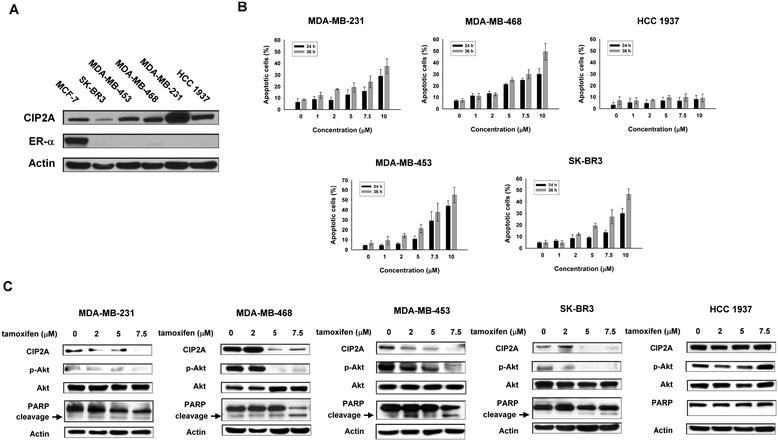
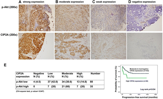
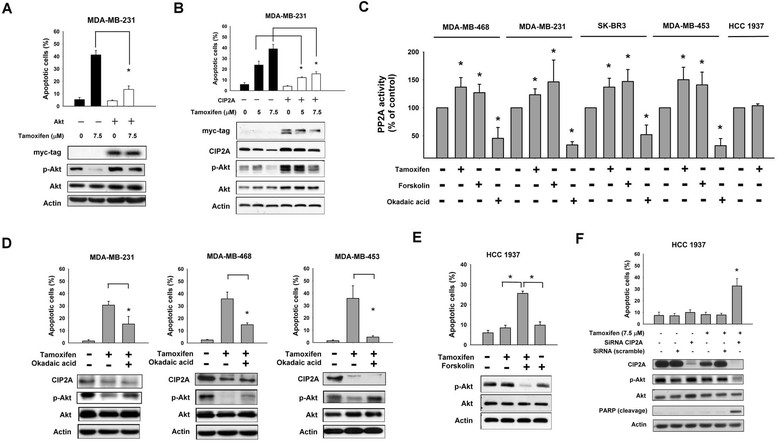
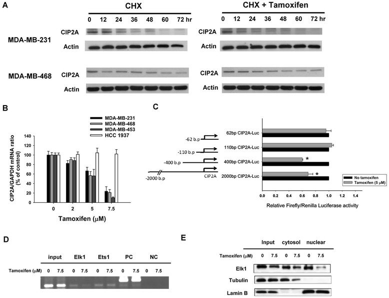
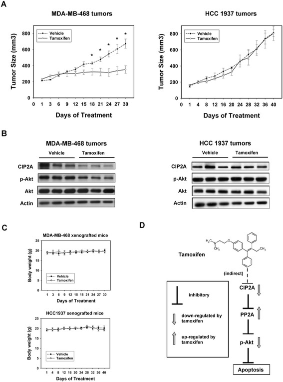
Tamoxifen induced significant apoptosis in MDA-MB-231, MDA-MB-468, MDA-MB-453 and SK-BR-3 cells, but not in HCC-1937 cells. Tamoxifen-induced apoptosis was associated with inhibition of cancerous inhibitor of protein phosphatase 2A (CIP2A) and phospho-Akt (p-Akt) in a dose-dependent manner. Ectopic expression of either CIP2A or Akt protected MDA-MB-231 cells from tamoxifen-induced apoptosis. In addition, tamoxifen increased protein phosphatase 2A (PP2A) activity, and tamoxifen-induced apoptosis was attenuated by the PP2A antagonist okadaic acid in the sensitive cell lines, but not in resistant HCC-1937 cells. Moreover, silencing CIP2A by small interfering RNA sensitized HCC-1937 cells to tamoxifen-induced apoptosis. Furthermore, tamoxifen regulated CIP2A protein expression by downregulating CIP2A mRNA. Importantly, tamoxifen inhibited the in vivo growth of MDA-MB-468 xenograft tumors in association with CIP2A downregulation, whereas tamoxifen had no significant effect on CIP2A expression and anti-tumor growth in HCC-1937 tumors.
Inhibition of CIP2A determines the effects of tamoxifen-induced apoptosis in ER-negative breast cancer cells. Our data suggest a novel "off-target" mechanism of tamoxifen and suggest that CIP2A/PP2A/p-Akt signaling may be a feasible anti-cancer pathway.
他莫昔芬是一种选择性雌激素受体(ER)调节剂,可能通过ER拮抗作用以外的机制影响癌细胞存活。在本研究中,我们测试了他莫昔芬在一组ER阴性乳腺癌细胞系中的疗效,并研究了其作用机制。
总共使用了五种ER阴性乳腺癌细胞系(HCC-1937、MDA-MB-231、MDA-MB-468、MDA-MB-453和SK-BR-3)进行体外研究。通过流式细胞术和蛋白质印迹分析检测细胞凋亡。通过蛋白质印迹分析评估细胞中的信号转导通路。在异种移植裸鼠中测试了他莫昔芬的体内疗效。
他莫昔芬在MDA-MB-231、MDA-MB-468、MDA-MB-453和SK-BR-3细胞中诱导了显著的凋亡,但在HCC-1937细胞中未诱导凋亡。他莫昔芬诱导的凋亡与剂量依赖性地抑制蛋白磷酸酶2A(CIP2A)癌性抑制剂和磷酸化Akt(p-Akt)有关。CIP2A或Akt的异位表达保护MDA-MB-231细胞免受他莫昔芬诱导的凋亡。此外,他莫昔芬增加了蛋白磷酸酶2A(PP2A)的活性,在敏感细胞系中,他莫昔芬诱导的凋亡被PP2A拮抗剂冈田酸减弱,但在耐药的HCC-1937细胞中未减弱。此外,通过小干扰RNA沉默CIP2A使HCC-1937细胞对他莫昔芬诱导的凋亡敏感。此外,他莫昔芬通过下调CIP2A mRNA来调节CIP2A蛋白表达。重要的是,他莫昔芬与CIP2A下调相关地抑制了MDA-MB-468异种移植肿瘤的体内生长,而他莫昔芬对HCC-1937肿瘤中的CIP2A表达和抗肿瘤生长没有显著影响。
抑制CIP2A决定了他莫昔芬诱导ER阴性乳腺癌细胞凋亡的作用。我们的数据提示了他莫昔芬一种新的“脱靶”机制,并提示CIP2A/PP2A/p-Akt信号通路可能是一条可行的抗癌途径。