Land Sander, Niederer Steven A, Louch William E, Røe Åsmund T, Aronsen Jan Magnus, Stuckey Daniel J, Sikkel Markus B, Tranter Matthew H, Lyon Alexander R, Harding Sian E, Smith Nicolas P
Department of Biomedical Engineering, King's College London, London, United Kingdom;
Institute for Experimental Medical Research, Oslo University Hospital Ullevål, Oslo, Norway; KG Jebsen Cardiac Research Center and Center for Heart Failure Research, University of Oslo, Oslo, Norway;
Am J Physiol Heart Circ Physiol. 2014 Nov 15;307(10):H1487-96. doi: 10.1152/ajpheart.00443.2014. Epub 2014 Sep 19.
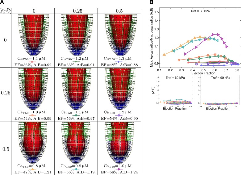
In Takotsubo cardiomyopathy, the left ventricle shows apical ballooning combined with basal hypercontractility. Both clinical observations in humans and recent experimental work on isolated rat ventricular myocytes suggest the dominant mechanisms of this syndrome are related to acute catecholamine overload. However, relating observed differences in single cells to the capacity of such alterations to result in the extreme changes in ventricular shape seen in Takotsubo syndrome is difficult. By using a computational model of the rat left ventricle, we investigate which mechanisms can give rise to the typical shape of the ventricle observed in this syndrome. Three potential dominant mechanisms related to effects of β-adrenergic stimulation were considered: apical-basal variation of calcium transients due to differences in L-type and sarco(endo)plasmic reticulum Ca(2+)-ATPase activation, apical-basal variation of calcium sensitivity due to differences in troponin I phosphorylation, and apical-basal variation in maximal active tension due to, e.g., the negative inotropic effects of p38 MAPK. Furthermore, we investigated the interaction of these spatial variations in the presence of a failing Frank-Starling mechanism. We conclude that a large portion of the apex needs to be affected by severe changes in calcium regulation or contractile function to result in apical ballooning, and smooth linear variation from apex to base is unlikely to result in the typical ventricular shape observed in this syndrome. A failing Frank-Starling mechanism significantly increases apical ballooning at end systole and may be an important additional factor underpinning Takotsubo syndrome.
在应激性心肌病中,左心室表现为心尖部膨出并伴有基底部过度收缩。人类的临床观察以及近期对离体大鼠心室肌细胞的实验研究均表明,该综合征的主要机制与急性儿茶酚胺超载有关。然而,将单个细胞中观察到的差异与这种改变导致应激性心肌病中所见心室形状极端变化的能力联系起来却很困难。通过使用大鼠左心室的计算模型,我们研究了哪些机制可能导致该综合征中观察到的典型心室形状。我们考虑了与β-肾上腺素能刺激效应相关的三种潜在主要机制:由于L型和肌浆网Ca(2+)-ATP酶激活差异导致的钙瞬变的尖基部差异、由于肌钙蛋白I磷酸化差异导致的钙敏感性的尖基部差异以及例如p38丝裂原活化蛋白激酶的负性肌力作用导致的最大主动张力的尖基部差异。此外,我们研究了在Frank-Starling机制衰竭的情况下这些空间差异的相互作用。我们得出结论,心尖的很大一部分需要受到钙调节或收缩功能的严重变化影响才能导致心尖部膨出,并且从心尖到基部的平滑线性变化不太可能导致该综合征中观察到的典型心室形状。Frank-Starling机制衰竭会显著增加收缩末期的心尖部膨出,可能是应激性心肌病的一个重要附加因素。