Grace P M, Ramos K M, Rodgers K M, Wang X, Hutchinson M R, Lewis M T, Morgan K N, Kroll J L, Taylor F R, Strand K A, Zhang Y, Berkelhammer D, Huey M G, Greene L I, Cochran T A, Yin H, Barth D S, Johnson K W, Rice K C, Maier S F, Watkins L R
Department of Psychology and the Center for Neuroscience, University of Colorado Boulder, Boulder, CO, USA.
Department of Psychology and the Center for Neuroscience, University of Colorado Boulder, Boulder, CO, USA.
Neuroscience. 2014 Nov 7;280:299-317. doi: 10.1016/j.neuroscience.2014.09.020. Epub 2014 Sep 18.
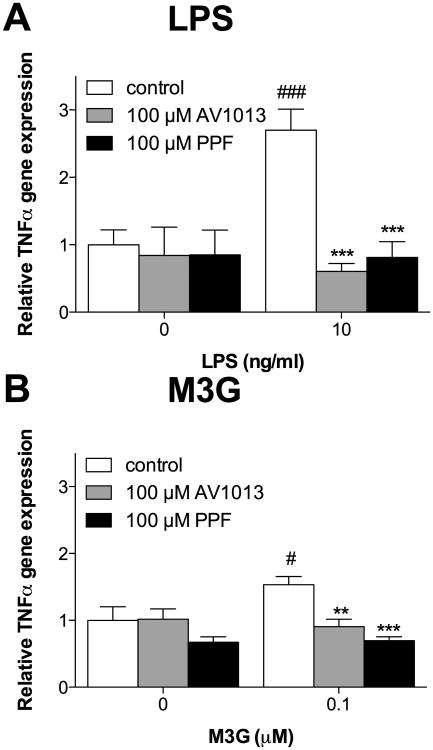
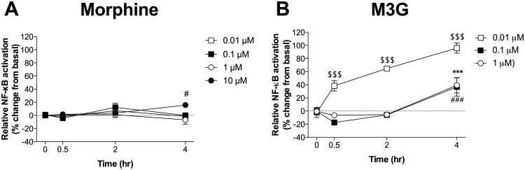
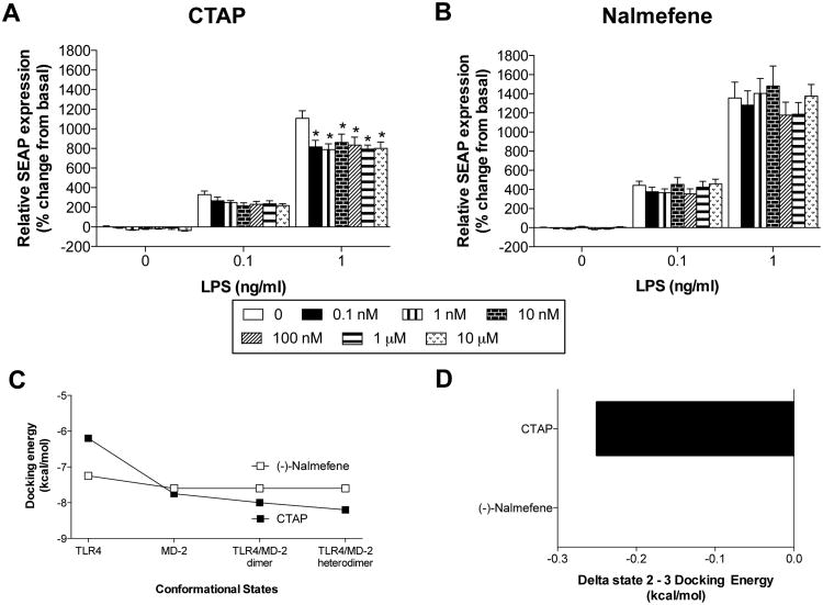
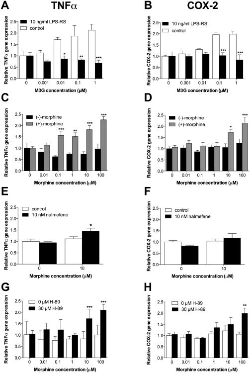
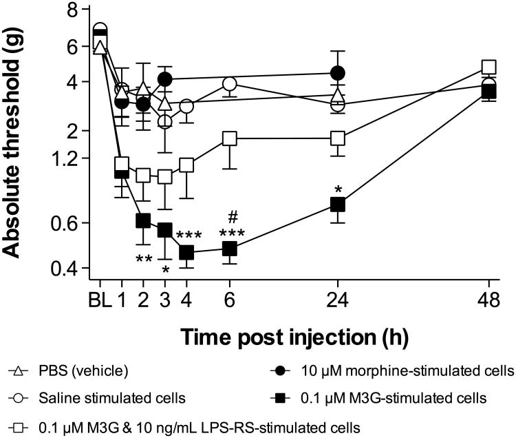
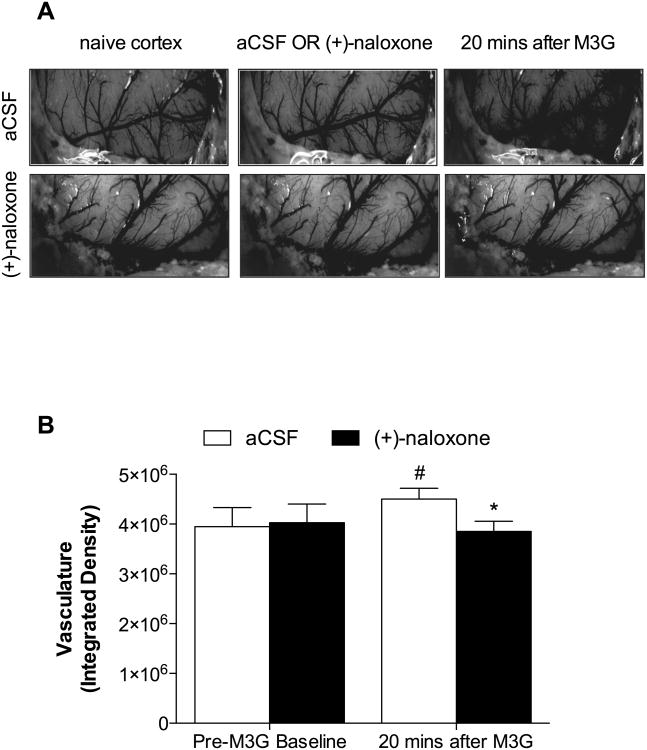
CNS immune signaling contributes to deleterious opioid effects including hyperalgesia, tolerance, reward, and dependence/withdrawal. Such effects are mediated by opioid signaling at toll-like receptor 4 (TLR4), presumptively of glial origin. Whether CNS endothelial cells express TLR4 is controversial. If so, they would be well positioned for activation by blood-borne opioids, contributing to opioid-induced pro-inflammatory responses. These studies examined adult primary rat CNS endothelial cell responses to (-)-morphine or its mu opioid receptor (MOR)-inactive metabolite morphine-3-glucuronide (M3G), both known TLR4 agonists. We demonstrate that adult rat CNS endothelial cells express functional TLR4. M3G activated nuclear factor kappaB (NF-κB), increased tumor necrosis factor-α (TNFα) and cyclooxygenase-2 (COX2) mRNAs, and released prostaglandin E2 (PGE2) from these cells. (-)-Morphine-induced upregulation of TNFα mRNA and PGE2 release were unmasked by pre-treatment with nalmefene, a MOR antagonist without TLR4 activity (unlike CTAP, shown to have both MOR- and TLR4-activity), suggestive of an interplay between MOR and TLR4 co-activation by (-)-morphine. In support, MOR-dependent Protein Kinase A (PKA) opposed TLR4 signaling, as PKA inhibition (H-89) also unmasked (-)-morphine-induced TNFα and COX2 mRNA upregulation. Intrathecal injection of CNS endothelial cells, stimulated in vitro with M3G, produced TLR4-dependent tactile allodynia. Further, cortical suffusion with M3G in vivo induced TLR4-dependent vasodilation. Finally, endothelial cell TLR4 activation by lipopolysaccharide and/or M3G was blocked by the glial inhibitors AV1013 and propentofylline, demonstrating endothelial cells as a new target of such drugs. These data indicate that (-)-morphine and M3G can activate CNS endothelial cells via TLR4, inducing proinflammatory, biochemical, morphological, and behavioral sequelae. CNS endothelial cells may have previously unanticipated roles in opioid-induced effects, in phenomena blocked by presumptive glial inhibitors, as well as TLR4-mediated phenomena more broadly.
中枢神经系统免疫信号传导会导致有害的阿片类药物效应,包括痛觉过敏、耐受性、奖赏和依赖/戒断。这些效应由 Toll 样受体 4(TLR4)上的阿片类信号传导介导,推测其起源于神经胶质细胞。中枢神经系统内皮细胞是否表达 TLR4 存在争议。如果表达,它们将易于被血源性阿片类药物激活,从而导致阿片类药物诱导的促炎反应。这些研究检测了成年原代大鼠中枢神经系统内皮细胞对(-)-吗啡或其μ阿片受体(MOR)无活性代谢物吗啡-3-葡萄糖醛酸苷(M3G)的反应,这两种物质都是已知的 TLR4 激动剂。我们证明成年大鼠中枢神经系统内皮细胞表达功能性 TLR4。M3G 激活核因子κB(NF-κB),增加肿瘤坏死因子-α(TNFα)和环氧化酶-2(COX2)的 mRNA 水平,并从这些细胞中释放前列腺素 E2(PGE2)。(-)-吗啡诱导的 TNFα mRNA 上调和 PGE2 释放可被纳美芬预处理所揭示,纳美芬是一种没有 TLR4 活性的 MOR 拮抗剂(与显示同时具有 MOR 和 TLR4 活性的 CTAP 不同),这表明(-)-吗啡在 MOR 和 TLR4 共同激活之间存在相互作用。此外,MOR 依赖性蛋白激酶 A(PKA)对抗 TLR4 信号传导,因为 PKA 抑制(H-89)也揭示了(-)-吗啡诱导的 TNFα 和 COX2 mRNA 上调。鞘内注射经 M3G 体外刺激的中枢神经系统内皮细胞会产生 TLR4 依赖性触觉异常性疼痛。此外,体内向皮质灌注 M3G 会诱导 TLR4 依赖性血管舒张。最后,脂多糖和/或 M3G 对内皮细胞 TLR4 的激活被神经胶质细胞抑制剂 AV1013 和丙戊茶碱阻断,这表明内皮细胞是这类药物的新靶点。这些数据表明(-)-吗啡和 M3G 可通过 TLR4 激活中枢神经系统内皮细胞,诱导促炎、生化、形态和行为后遗症。中枢神经系统内皮细胞可能在阿片类药物诱导的效应、推测的神经胶质细胞抑制剂所阻断的现象以及更广泛的 TLR4 介导的现象中发挥先前未被预料到的作用。