Acosta Sandra A, Tajiri Naoki, de la Pena Ike, Bastawrous Marina, Sanberg Paul R, Kaneko Yuji, Borlongan Cesar V
Center of Excellence for Aging and Brain Repair, Department of Neurosurgery and Brain Repair, University of South Florida College of Medicine, Tampa, Florida.
J Cell Physiol. 2015 May;230(5):1024-32. doi: 10.1002/jcp.24830.
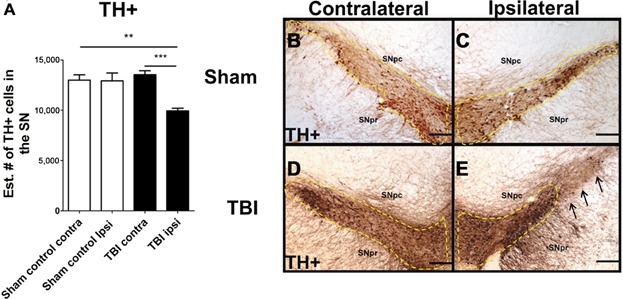
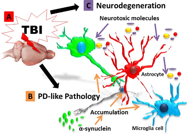
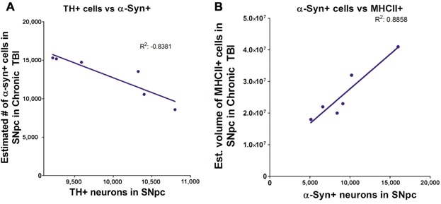
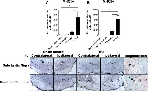
The long-term consequences of traumatic brain injury (TBI) are closely associated with the development of histopathological deficits. Notably, TBI may predispose long-term survivors to age-related neurodegenerative diseases, such as Parkinson's disease (PD), which is characterized by a gradual degeneration of the nigrostriatal dopaminergic neurons. However, preclinical studies on the pathophysiological changes in substantia nigra (SN) after chronic TBI are lacking. In the present in vivo study, we examined the pathological link between PD-associated dopaminergic neuronal loss and chronic TBI. Sixty days post-TBI, rats were euthanized and brain tissues harvested. Immunostaining was performed using tyrosine hydroxylase (TH), an enzyme required for the synthesis of dopamine in neurons, α-synuclein, a presynaptic protein that plays a role in synaptic vesicle recycling, and major histocompatibility complex II (MHCII), a protein found in antigen presenting cells such as inflammatory microglia cells, all key players in PD pathology. Unbiased stereology analyses revealed significant decrease of TH-positive expression in the surviving dopaminergic neurons of the SN pars compacta (SNpc) relative to sham control. In parallel, increased α-synuclein accumulation was detected in the ipsilateral SN compared to the contralateral SN in TBI animals or sham control. In addition, exacerbation of MHCII+ cells was recognized in the SN and cerebral peduncle ipsilateral to injury relative to contralateral side and sham control. These results suggest α-synuclein as a pathological link between chronic effects of TBI and PD symptoms as evidenced by significant overexpression and abnormal accumulation of α-synuclein in inflammation-infiltrated SN of rats exposed to chronic TBI.
创伤性脑损伤(TBI)的长期后果与组织病理学缺陷的发展密切相关。值得注意的是,TBI可能使长期幸存者易患与年龄相关的神经退行性疾病,如帕金森病(PD),其特征是黑质纹状体多巴胺能神经元逐渐退化。然而,目前缺乏关于慢性TBI后黑质(SN)病理生理变化的临床前研究。在本体内研究中,我们研究了PD相关多巴胺能神经元丢失与慢性TBI之间的病理联系。TBI后60天,对大鼠实施安乐死并采集脑组织。使用酪氨酸羟化酶(TH,神经元中多巴胺合成所需的一种酶)、α-突触核蛋白(一种在前突触蛋白中起作用的突触囊泡循环蛋白)和主要组织相容性复合体II(MHCII,一种在抗原呈递细胞如炎性小胶质细胞中发现的蛋白质,这些都是PD病理学中的关键因素)进行免疫染色。无偏倚的体视学分析显示,相对于假手术对照组,致密部黑质(SNpc)中存活的多巴胺能神经元中TH阳性表达显著降低。同时,与TBI动物或假手术对照组的对侧SN相比,同侧SN中α-突触核蛋白积累增加。此外,相对于对侧和假手术对照组,损伤同侧的SN和脑桥中MHCII +细胞增多。这些结果表明,α-突触核蛋白是TBI慢性效应与PD症状之间的病理联系,这一点可通过慢性TBI大鼠炎症浸润的SN中α-突触核蛋白的显著过表达和异常积累得到证明。