Aizawa Naoko, Kunikata Hiroshi, Shiga Yukihiro, Yokoyama Yu, Omodaka Kazuko, Nakazawa Toru
Department of Ophthalmology, Tohoku University Graduate School of Medicine, 1-1 Seiryo-machi, Aoba-ku, Sendai 980-8574, Japan.
BMC Ophthalmol. 2014 Sep 24;14:113. doi: 10.1186/1471-2415-14-113.
It is difficult to identify glaucoma in myopic eyes because the configuration of the optic disc varies; yet it is important clinically. Here, we used laser speckle flowgraphy (LSFG) to measure mean blur rate (MBR), representing optic disc microcirculation, and assessed its ability to identify glaucoma in eyes with myopic optic discs.
129 eyes (normal disc: 21 eyes; myopic disc: 108 eyes) were enrolled. The eyes were classified as normal or mildly, moderately, or severely glaucomatous with standard automated perimetry (SAP). We determined the relationship between optic nerve head (ONH) MBR, measured with LSFG, mean deviation (MD), measured with SAP, and circumpapillary retinal nerve fiber layer thickness (cpRNFLT), measured with optical coherence tomography (OCT).
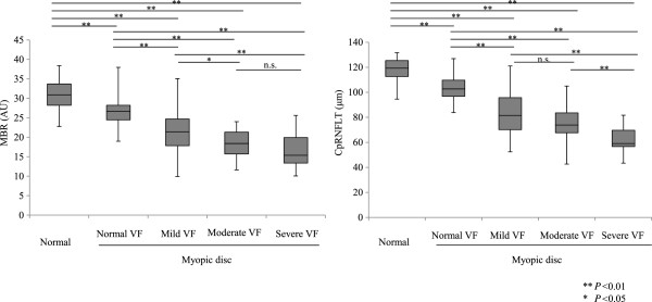
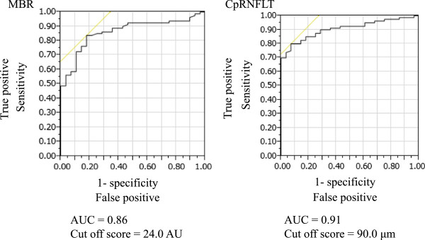
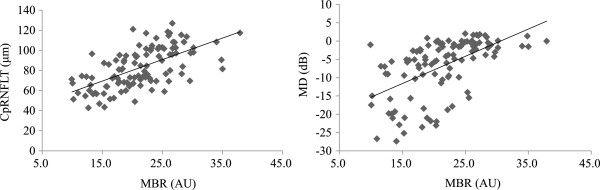
ONH MBR and cpRNFLT decreased significantly with the severity of glaucoma. MBR was significantly correlated with cpRNFLT and MD (r =0.65 and r =0.63, respectively). A multiple regression analysis revealed that MBR and cpRNFLT were independent factors indicating glaucoma severity. A logistic regression analysis revealed that MBR and cpRNFLT were also independent factors indicating the presence of glaucoma. In a receiver operating characteristic (ROC) analysis, MBR and cpRNFLT could both differentiate between normal and glaucomatous eyes (MBR area under the ROC curve: 0.86, with a cut-off score of 24.0 AU).
These results suggest that in addition to cpRNFLT, non-invasive and objective LSFG measurements of MBR may enable the identification of glaucoma and the classification of its severity in eyes with myopic optic discs.
由于近视眼视盘形态各异,因此很难识别其中的青光眼;然而在临床上这很重要。在此,我们使用激光散斑血流图(LSFG)测量代表视盘微循环的平均模糊率(MBR),并评估其识别具有近视性视盘的眼睛中青光眼的能力。
纳入129只眼睛(正常视盘:21只眼;近视视盘:108只眼)。通过标准自动视野计(SAP)将眼睛分类为正常、轻度、中度或重度青光眼。我们确定了用LSFG测量的视神经乳头(ONH)MBR、用SAP测量的平均偏差(MD)以及用光学相干断层扫描(OCT)测量的视盘周围视网膜神经纤维层厚度(cpRNFLT)之间的关系。
随着青光眼严重程度增加,ONH MBR和cpRNFLT显著降低。MBR与cpRNFLT和MD显著相关(分别为r = 0.65和r = 0.63)。多元回归分析显示,MBR和cpRNFLT是表明青光眼严重程度的独立因素。逻辑回归分析显示,MBR和cpRNFLT也是表明青光眼存在的独立因素。在受试者工作特征(ROC)分析中,MBR和cpRNFLT均可区分正常眼和青光眼眼(ROC曲线下面积:MBR为0.86,临界值为24.0 AU)。
这些结果表明,除了cpRNFLT之外,对MBR进行非侵入性和客观性的LSFG测量可能有助于识别具有近视性视盘的眼睛中的青光眼并对其严重程度进行分级。