Drogoszewska Barbara, Chomik Piotr, Polcyn Adam, Michcik Adam
Chair and Department of Oral and Maxillofacial Surgery, Medical University of Gdansk, Poland. Head of Department: Prof. Adam Włodarkiewicz MD, DMD, PhD ; Department of Otolaryngology, Ward of Maxillofacial Surgery, University Clinical Center, Gdansk, Poland. Head of Department: Prof. Adam Włodarkiewicz MD, DMD, PhD.
Department of Otolaryngology, Ward of Maxillofacial Surgery, University Clinical Center, Gdansk, Poland. Head of Department: Prof. Adam Włodarkiewicz MD, DMD, PhD.
Postepy Dermatol Alergol. 2014 Aug;31(4):222-8. doi: 10.5114/pdia.2014.40926. Epub 2014 Sep 8.
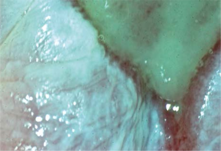
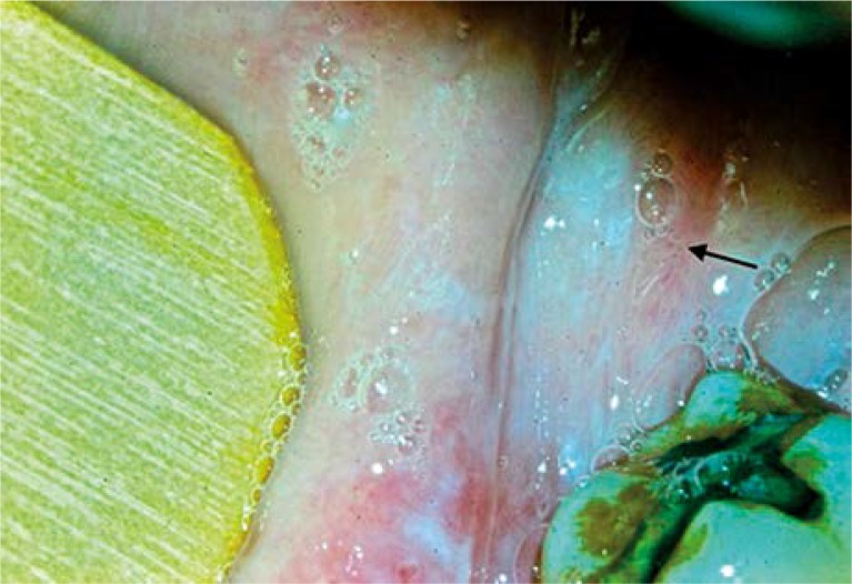
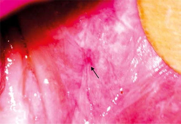
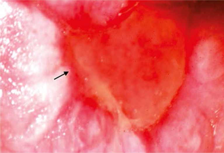
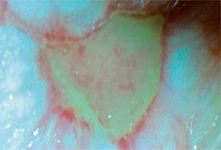
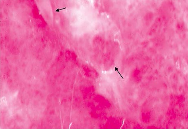
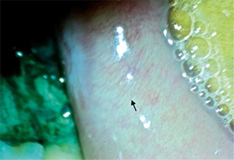
Direct oral microscopy is a novel, non-invasive diagnostic technique that aids clinical examination of the oral cavity. The basic principles of this method derive from colposcopy and dermoscopy. The principle is to reveal precancerous lesions of oral mucosae in their subclinical phase in order to begin their treatment as early as possible and prevent malignant transformation. Oral lichen planus (OLP) is an autoimmune, inflammatory, chronic disease affecting oral mucous membranes. Buccal mucosae are most often affected.
To describe the in vivo picture of erosive OLP in direct oral microscopy in terms of the pattern and density of subepithelial blood vessels, surface texture, color, transparency and borders of the lesions. The study also demonstrates the utility of the method in the selection of the most appropriate biopsy site.
A total of 30 patients with erosive OLP were examined. Clinical examination of the oral cavity with the naked eye was performed, followed by direct oral microscopy. The most appropriate biopsy sites based on both examinations were chosen for every individual and biopsies were taken for histopathological evaluation.
Biopsies obtained based on direct oral microscopy revealed dysplasia in 16 patients (53.3%). Biopsies obtained based on clinical examination with the naked eye revealed dysplasia in 3 cases (10%).
Direct oral microscopy makes it possible to obtain a repeated picture of erosive OLP and constitutes an alternative to the clinical examination with the naked eye in election of the most appropriate biopsy site. Thus, introduction of the most accurate and early therapy is possible.
直接口腔显微镜检查是一种新型的非侵入性诊断技术,有助于口腔的临床检查。该方法的基本原理源自阴道镜检查和皮肤镜检查。其原理是揭示口腔黏膜癌前病变的亚临床阶段,以便尽早开始治疗并预防恶性转化。口腔扁平苔藓(OLP)是一种影响口腔黏膜的自身免疫性、炎症性慢性疾病。颊黏膜最常受累。
从上皮下血管的形态和密度、表面纹理、颜色、透明度及病变边界等方面描述直接口腔显微镜检查下糜烂性OLP的活体图像。本研究还证明了该方法在选择最合适活检部位方面的实用性。
共检查了30例糜烂性OLP患者。先进行肉眼口腔临床检查,然后进行直接口腔显微镜检查。根据这两种检查为每个患者选择最合适的活检部位,并进行活检以进行组织病理学评估。
基于直接口腔显微镜检查获取的活检标本显示16例患者(53.3%)存在发育异常。基于肉眼临床检查获取的活检标本显示3例(10%)存在发育异常。
直接口腔显微镜检查能够获取糜烂性OLP的重复图像,并且在选择最合适的活检部位方面可替代肉眼临床检查。因此,可以采用最准确的早期治疗方法。