Gruda Yuliia, Albrecht Marius, Buckova Michaela, Haim Dominik, Lauer Guenter, Koch Edmund, Joehrens Korinna, Schnabel Christian, Golde Jonas, Li Jiawen, McLaughlin Robert A, Walther Julia
Carl Gustav Carus Faculty of Medicine, Department of Medical Physics and Biomedical Engineering, TU Dresden, Fetscherstraße 74, 01307 Dresden, Germany.
Carl Gustav Carus Faculty of Medicine, Institute of Pathology, TU Dresden, Fetscherstraße 74, 01307 Dresden, Germany.
Diagnostics (Basel). 2023 Aug 10;13(16):2642. doi: 10.3390/diagnostics13162642.
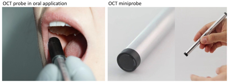
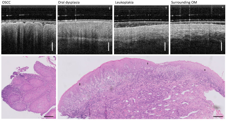
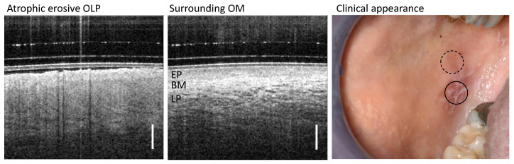
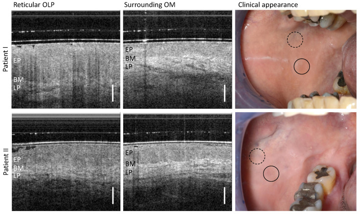
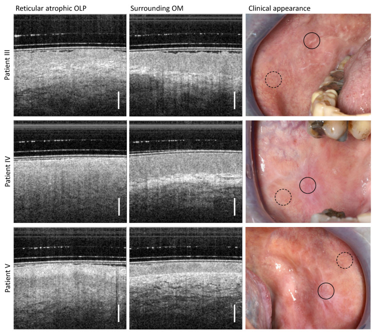
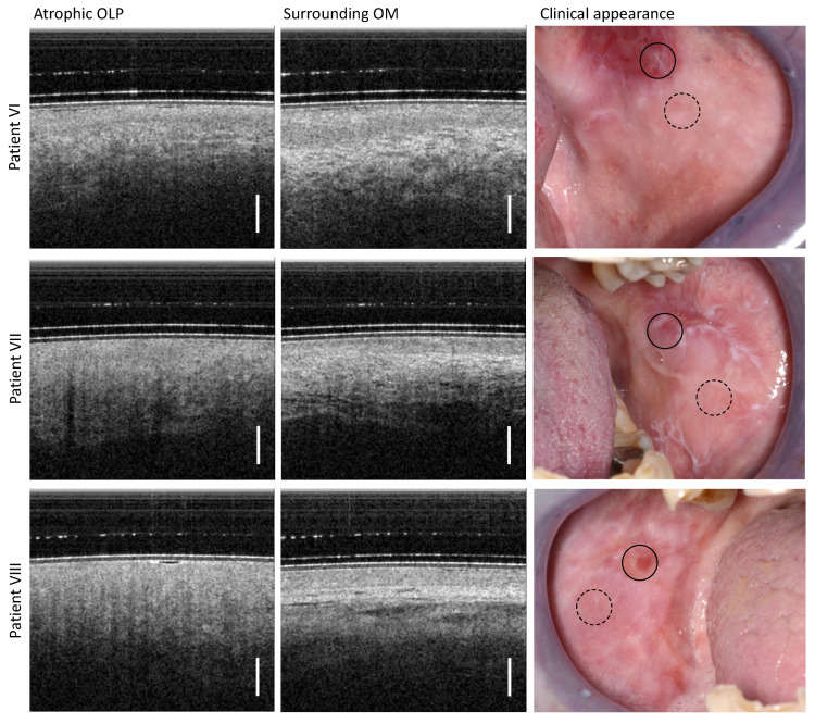
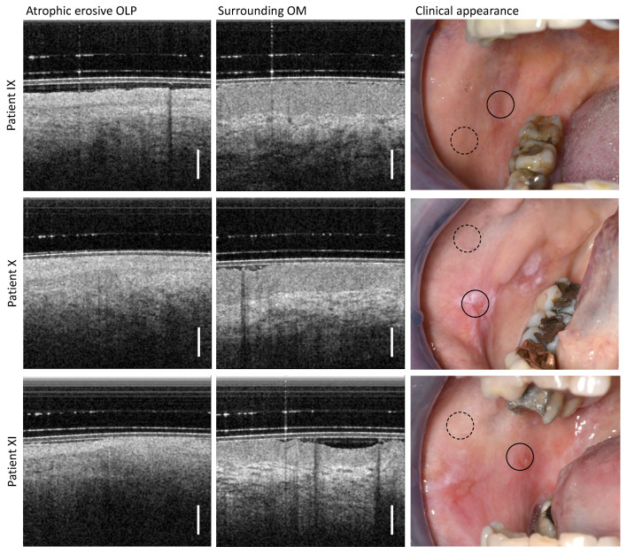
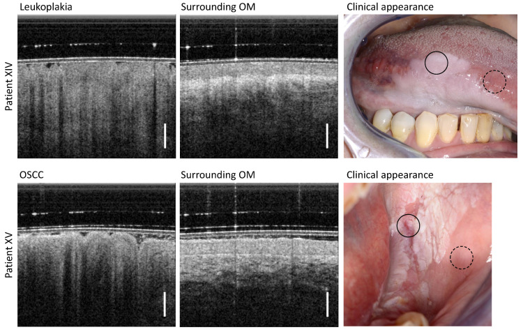
Malignant transformation of oral lichen planus (OLP) into oral squamous cell carcinoma is considered as one of the most serious complications of OLP. For the early detection of oral cancer in OLP follow-up, accurate localization of the OLP center is still difficult but often required for confirmatory biopsy with histopathological examination. Optical coherence tomography (OCT) offers the potential for more reliable biopsy sampling in the oral cavity as it is capable of non-invasively imaging the degenerated oral layer structure. In this case-series study with 15 patients, features of clinically classified forms of OLP in OCT cross-sections were registered and correlated with available histologic sections. Besides patients with reticular, atrophic, erosive and plaque-like OLP, two patients with leukoplakia were included for differentiation. The results show that OCT yields information about the epithelial surface, thickness and reflectivity, as well as the identifiability of the basement membrane and the vessel network, which could be used to complement the visual clinical appearance of OLP variants and allow a more accurate localization of the OLP center. This forms the basis for further studies on OCT-assisted non-invasive clinical classification of OLP, with the aim of enabling decision support for biopsy sampling in the future.
口腔扁平苔藓(OLP)恶变成为口腔鳞状细胞癌被认为是OLP最严重的并发症之一。在OLP随访中进行口腔癌的早期检测时,OLP中心的准确定位仍然困难,但在进行组织病理学检查的确诊活检时却常常需要。光学相干断层扫描(OCT)能够对退化的口腔层结构进行无创成像,为在口腔中进行更可靠的活检取样提供了可能。在这项针对15名患者的病例系列研究中,记录了OCT横截面中临床分类的OLP形式的特征,并将其与现有的组织学切片进行关联。除了网状、萎缩性、糜烂性和斑块状OLP患者外,还纳入了两名白斑患者以进行鉴别。结果表明,OCT可提供有关上皮表面、厚度和反射率的信息,以及基底膜和血管网络的可识别性,这些信息可用于补充OLP变体的视觉临床外观,并实现OLP中心的更准确定位。这为进一步研究OCT辅助的OLP无创临床分类奠定了基础,目的是在未来为活检取样提供决策支持。