文献检索文档翻译深度研究
Suppr Zotero 插件Zotero 插件
邀请有礼套餐&价格历史记录

新学期,新优惠

限时优惠:9月1日-9月22日

30天高级会员仅需29元

1天体验卡首发特惠仅需5.99元

了解详情
不再提醒
插件&应用
Suppr Zotero 插件Zotero 插件浏览器插件Mac 客户端Windows 客户端微信小程序
高级版
套餐订阅购买积分包
AI 工具
文献检索文档翻译深度研究
关于我们
关于 Suppr公司介绍联系我们用户协议隐私条款
关注我们

Suppr 超能文献

核心技术专利:CN118964589B侵权必究
粤ICP备2023148730 号-1Suppr @ 2025

关节中的细胞因子谱取决于病理状况,但在滑液、软骨组织和培养的软骨细胞之间存在差异。

Cytokine profiles in the joint depend on pathology, but are different between synovial fluid, cartilage tissue and cultured chondrocytes.

作者信息

Tsuchida Anika I, Beekhuizen Michiel, 't Hart Marieke C, Radstake Timothy R D J, Dhert Wouter J A, Saris Daniel B F, van Osch Gerjo J V M, Creemers Laura B

出版信息

Arthritis Res Ther. 2014 Sep 26;16(5):441. doi: 10.1186/s13075-014-0441-0.


DOI:10.1186/s13075-014-0441-0
PMID:25256035
原文链接:https://pmc.ncbi.nlm.nih.gov/articles/PMC4201683/
Abstract

INTRODUCTION: This study aimed to evaluate whether profiles of several soluble mediators in synovial fluid and cartilage tissue are pathology-dependent and how their production is related to in vitro tissue formation by chondrocytes from diseased and healthy tissue. METHODS: 
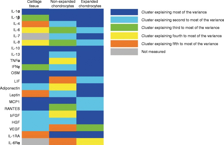
Samples were obtained from donors without joint pathology (n = 39), with focal defects (n = 65) and osteoarthritis (n = 61). A multiplex bead assay (Luminex) was performed measuring up to 21 cytokines: Interleukin (IL)-1α, IL-1β, IL-1RA, IL-4, IL-6, IL-6Rα, IL-7, IL-8, IL-10, IL-13, tumor necrosis factor (TNF)α, Interferon (IFN)γ, oncostatin M (OSM), leukemia inhibitory factor (LIF), adiponectin, leptin, monocyte chemotactic factor (MCP)1, RANTES, basic fibroblast growth factor (bFGF), hepatocyte growth factor (HGF), vascular growth factor (VEGF). RESULTS: 
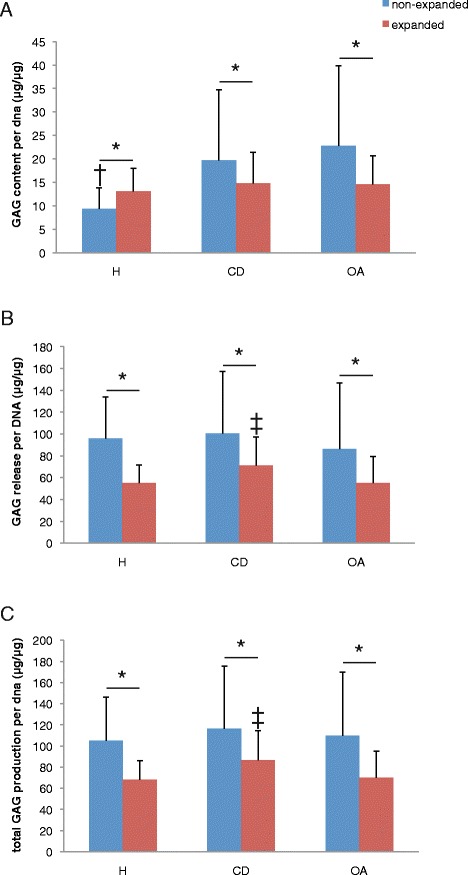
In synovial fluid of patients with cartilage pathology, IL-6, IL-13, IFNγ and OSM levels were higher than in donors without joint pathology (P ≤ 0.001). IL-13, IFNγ and OSM were also different between donors with cartilage defects and OA (P < 0.05). In cartilage tissue from debrided defects, VEGF was higher than in non-pathological or osteoarthritic joints (P ≤ 0.001). IL-1α, IL-6, TNFα and OSM concentrations (in ng/ml) were markedly higher in cartilage tissue than in synovial fluid (P <0.01). Culture of chondrocytes generally led to a massive induction of most cytokines (P < 0.001). Although the release of inflammatory cytokines was also here dependent on the pathological condition (P < 0.001) the actual profiles were different from tissue or synovial fluid and between non-expanded and expanded chondrocytes. Cartilage formation was lower by healthy unexpanded chondrocytes than by osteoarthritic or defect chondrocytes. CONCLUSIONS: Several pro-inflammatory, pro-angiogenic and pro-repair cytokines were elevated in joints with symptomatic cartilage defects and/or osteoarthritis, although different cytokines were elevated in synovial fluid compared to tissue or cells. Hence a clear molecular profile was evident dependent on disease status of the joint, which however changed in composition depending on the biological sample analysed. These alterations did not affect in vitro tissue formation with these chondrocytes, as this was at least as effective or even better compared to healthy chondrocytes.

摘要

引言:本研究旨在评估滑液和软骨组织中几种可溶性介质的概况是否与病理状况相关,以及它们的产生与来自患病和健康组织的软骨细胞的体外组织形成有何关系。 方法:样本取自无关节病变的供体(n = 39)、有局灶性缺损的供体(n = 65)和骨关节炎患者(n = 61)。采用多重微珠检测法(Luminex)检测多达21种细胞因子:白细胞介素(IL)-1α、IL-1β、IL-1受体拮抗剂(IL-1RA)、IL-4、IL-6、IL-6受体α(IL-6Rα)、IL-7、IL-8、IL-10、IL-13、肿瘤坏死因子(TNF)α、干扰素(IFN)γ、制瘤素M(OSM)、白血病抑制因子(LIF)、脂联素、瘦素、单核细胞趋化因子(MCP)1、调节激活正常T细胞表达和分泌因子(RANTES)、碱性成纤维细胞生长因子(bFGF)、肝细胞生长因子(HGF)、血管生长因子(VEGF)。 结果:在有软骨病变患者的滑液中,IL-6、IL-13、IFNγ和OSM水平高于无关节病变的供体(P≤0.001)。IL-13、IFNγ和OSM在有软骨缺损的供体与骨关节炎患者之间也存在差异(P<0.05)。在清创缺损的软骨组织中,VEGF高于非病理或骨关节炎关节(P≤0.001)。软骨组织中IL-1α、IL-6、TNFα和OSM的浓度(以ng/ml计)明显高于滑液(P<0.01)。软骨细胞培养通常会导致大多数细胞因子的大量诱导(P<0.001)。虽然炎症细胞因子的释放也取决于病理状况(P<0.001),但其实际概况与组织或滑液不同,且在未扩增和扩增的软骨细胞之间也不同。健康未扩增的软骨细胞形成的软骨比骨关节炎或缺损软骨细胞形成的软骨少。 结论:在有症状性软骨缺损和/或骨关节炎的关节中,几种促炎、促血管生成和促修复的细胞因子升高,尽管与组织或细胞相比,滑液中升高的细胞因子不同。因此,根据关节疾病状态明显有一个清晰的分子概况,然而其组成会根据所分析的生物样本而改变。这些改变并不影响这些软骨细胞的体外组织形成,因为与健康软骨细胞相比,这至少同样有效甚至更好。

https://cdn.ncbi.nlm.nih.gov/pmc/blobs/34db/4201683/73c75a09e30c/13075_2014_441_Fig4_HTML.jpg
https://cdn.ncbi.nlm.nih.gov/pmc/blobs/34db/4201683/bfa663e927d7/13075_2014_441_Fig1_HTML.jpg
https://cdn.ncbi.nlm.nih.gov/pmc/blobs/34db/4201683/e56301596aea/13075_2014_441_Fig2_HTML.jpg
https://cdn.ncbi.nlm.nih.gov/pmc/blobs/34db/4201683/5b0fdcb863da/13075_2014_441_Fig3_HTML.jpg
https://cdn.ncbi.nlm.nih.gov/pmc/blobs/34db/4201683/73c75a09e30c/13075_2014_441_Fig4_HTML.jpg

相似文献

[1]
Cytokine profiles in the joint depend on pathology, but are different between synovial fluid, cartilage tissue and cultured chondrocytes.

Arthritis Res Ther. 2014-9-26

[2]
Interleukin-6 is elevated in synovial fluid of patients with focal cartilage defects and stimulates cartilage matrix production in an in vitro regeneration model.

Arthritis Res Ther. 2012-12-3

[3]
Inhibition of oncostatin M in osteoarthritic synovial fluid enhances GAG production in osteoarthritic cartilage repair.

Eur Cell Mater. 2013-9-11

[4]
Synovial fluid levels of tumor necrosis factor alpha and oncostatin M correlate with levels of markers of the degradation of crosslinked collagen and cartilage aggrecan in rheumatoid arthritis but not in osteoarthritis.

Arthritis Rheum. 2000-2

[5]
Cytokine profile of autologous conditioned serum for treatment of osteoarthritis, in vitro effects on cartilage metabolism and intra-articular levels after injection.

Arthritis Res Ther. 2010-6-10

[6]
Interleukin-7 levels in synovial fluid increase with age and MMP-1 levels decrease with progression of osteoarthritis.

Acta Orthop. 2011-12-29

[7]
IL-40 is up-regulated in the synovial fluid and cartilage of osteoarthritis patients and contributes to the alteration of chondrocytes phenotype in vitro.

Arthritis Res Ther. 2024-7-30

[8]
Osteoarthritis synovial fluid activates pro-inflammatory cytokines in primary human chondrocytes.

Int Orthop. 2012-12-5

[9]
Anticytokine therapy for osteoarthritis: evidence to date.

Drugs Aging. 2010-2-1

[10]
The role of oncostatin M in animal and human connective tissue collagen turnover and its localization within the rheumatoid joint.

Arthritis Rheum. 1998-10

引用本文的文献

[1]
Targeting Cellular Senescence: Pathophysiology in Multisystem Age-Related Diseases.

Biomedicines. 2025-7-15

[2]
Isoginkgetin protects chondrocytes and inhibits osteoarthritis through NF-κB and P21 signaling pathway.

Mol Med. 2025-6-22

[3]
Atf3 + senescent chondrocytes mediate meniscus degeneration in aging.

Arthritis Res Ther. 2025-5-15

[4]
Clinical and Molecular Barriers to Understanding the Pathogenesis, Diagnosis, and Treatment of Complex Regional Pain Syndrome (CRPS).

Int J Mol Sci. 2025-3-11

[5]
Cellular and molecular mechanisms underlying obesity in degenerative spine and joint diseases.

Bone Res. 2024-12-11

[6]
Cross-talk of inflammation and cellular senescence: a new insight into the occurrence and progression of osteoarthritis.

Bone Res. 2024-12-3

[7]
Unraveling the complex interplay of sex, endocrinology, and inflammation in post-Injury articular cartilage breakdown through in silico modeling.

Sci Rep. 2024-11-19

[8]
Cellular Senescence: The Driving Force of Musculoskeletal Diseases.

Biomedicines. 2024-8-26

[9]
β-Hydroxybutyrate enhances chondrocyte mitophagy and reduces cartilage degeneration in osteoarthritis via the HCAR2/AMPK/PINK1/Parkin pathway.

Aging Cell. 2024-11

[10]
Disease-Modifying Effects of Lenvatinib, a Multiple Receptor Tyrosine Kinase Inhibitor, on Posttraumatic Osteoarthritis of the Knee.

Int J Mol Sci. 2024-6-13

本文引用的文献

[1]
Modeling of signaling pathways in chondrocytes based on phosphoproteomic and cytokine release data.

Osteoarthritis Cartilage. 2014-3

[2]
Chondrogenic potential of articular chondrocytes depends on their original location.

Tissue Eng Part A. 2013-11-14

[3]
Inhibition of oncostatin M in osteoarthritic synovial fluid enhances GAG production in osteoarthritic cartilage repair.

Eur Cell Mater. 2013-9-11

[4]
The effects of nonsteroidal anti-inflammatory drugs on clinical outcomes, synovial fluid cytokine concentration and signal transduction pathways in knee osteoarthritis. A randomized open label trial.

Osteoarthritis Cartilage. 2013-9

[5]
Intraarticular and systemic inflammatory profiles may identify patients with osteoarthritis.

J Rheumatol. 2013-5-15

[6]
Gene expression profiling of dedifferentiated human articular chondrocytes in monolayer culture.

Osteoarthritis Cartilage. 2013-1-30

[7]
Interleukin-4- and interleukin-13-mediated alternatively activated macrophages: roles in homeostasis and disease.

Annu Rev Immunol. 2013-1-3

[8]
Injection of vascular endothelial growth factor into knee joints induces osteoarthritis in mice.

Osteoarthritis Cartilage. 2012-12-19

[9]
Interleukin-6 is elevated in synovial fluid of patients with focal cartilage defects and stimulates cartilage matrix production in an in vitro regeneration model.

Arthritis Res Ther. 2012-12-3

[10]
Matrix-assisted autologous chondrocyte transplantation for cartilage regeneration in osteoarthritic knees: results and failures at midterm follow-up.

Am J Sports Med. 2012-10-25

文献AI研究员

20分钟写一篇综述,助力文献阅读效率提升50倍

立即体验

用中文搜PubMed

大模型驱动的PubMed中文搜索引擎

马上搜索

推荐工具

医学文档翻译智能文献检索