Mead Ben, Logan Ann, Berry Martin, Leadbeater Wendy, Scheven Ben A
Neurotrauma Research Group, Neurobiology Section, School of Clinical and Experimental Medicine, University of Birmingham, Birmingham, United Kingdom; School of Dentistry, University of Birmingham, Birmingham, United Kingdom.
Neurotrauma Research Group, Neurobiology Section, School of Clinical and Experimental Medicine, University of Birmingham, Birmingham, United Kingdom.
PLoS One. 2014 Oct 7;9(10):e109305. doi: 10.1371/journal.pone.0109305. eCollection 2014.
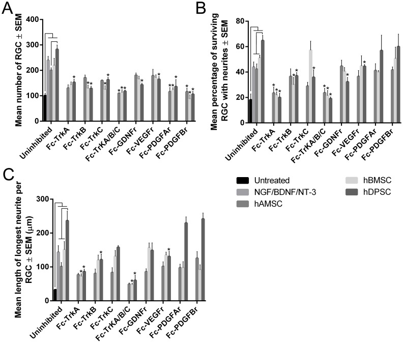
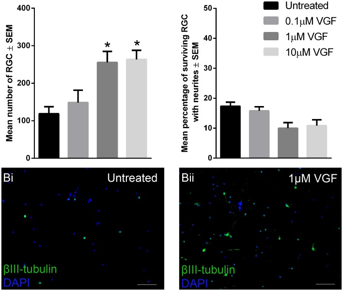
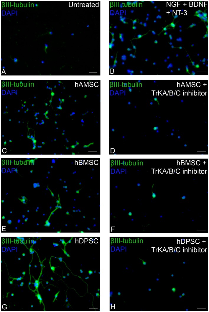
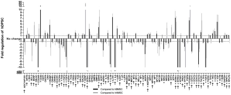
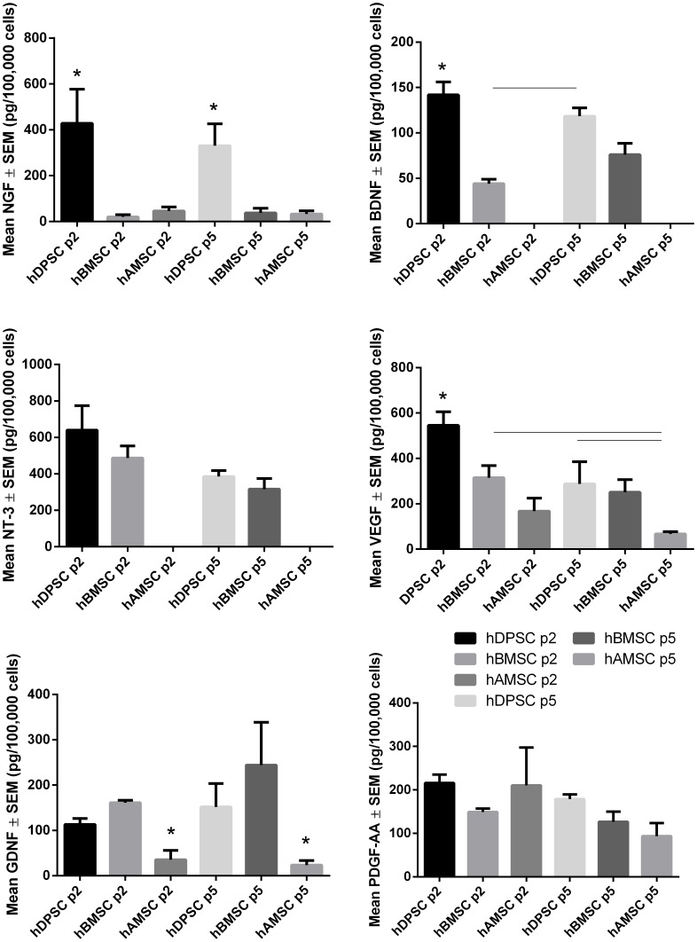
We have investigated and compared the neurotrophic activity of human dental pulp stem cells (hDPSC), human bone marrow-derived mesenchymal stem cells (hBMSC) and human adipose-derived stem cells (hAMSC) on axotomised adult rat retinal ganglion cells (RGC) in vitro in order to evaluate their therapeutic potential for neurodegenerative conditions of RGC. Using the transwell system, RGC survival and length/number of neurites were quantified in coculture with stem cells in the presence or absence of specific Fc-receptor inhibitors to determine the role of NGF, BDNF, NT-3, VEGF, GDNF, PDGF-AA and PDGF-AB/BB in stem cell-mediated RGC neuroprotection and neuritogenesis. Conditioned media, collected from cultured hDPSC/hBMSC/hAMSC, were assayed for the secreted growth factors detailed above using ELISA. PCR array determined the hDPSC, hBMSC and hAMSC expression of genes encoding 84 growth factors and receptors. The results demonstrated that hDPSC promoted significantly more neuroprotection and neuritogenesis of axotomised RGC than either hBMSC or hAMSC, an effect that was neutralized after the addition of specific Fc-receptor inhibitors. hDPSC secreted greater levels of various growth factors including NGF, BDNF and VEGF compared with hBMSC/hAMSC. The PCR array confirmed these findings and identified VGF as a novel potentially therapeutic hDPSC-derived neurotrophic factor (NTF) with significant RGC neuroprotective properties after coculture with axotomised RGC. In conclusion, hDPSC promoted significant multi-factorial paracrine-mediated RGC survival and neurite outgrowth and may be considered a potent and advantageous cell therapy for retinal nerve repair.
我们已经研究并比较了人牙髓干细胞(hDPSC)、人骨髓间充质干细胞(hBMSC)和人脂肪来源干细胞(hAMSC)对成年大鼠视网膜神经节细胞(RGC)轴突切断后体外的神经营养活性,以评估它们对RGC神经退行性疾病的治疗潜力。使用Transwell系统,在存在或不存在特异性Fc受体抑制剂的情况下,对与干细胞共培养的RGC存活率和神经突长度/数量进行定量,以确定NGF、BDNF、NT-3、VEGF、GDNF、PDGF-AA和PDGF-AB/BB在干细胞介导的RGC神经保护和神经突生成中的作用。从培养的hDPSC/hBMSC/hAMSC中收集的条件培养基,使用ELISA检测上述分泌的生长因子。PCR阵列测定了编码84种生长因子和受体的基因在hDPSC、hBMSC和hAMSC中的表达。结果表明,hDPSC比hBMSC或hAMSC显著促进更多轴突切断的RGC的神经保护和神经突生成,添加特异性Fc受体抑制剂后这种作用被中和。与hBMSC/hAMSC相比,hDPSC分泌更高水平的各种生长因子,包括NGF、BDNF和VEGF。PCR阵列证实了这些发现,并确定VGF是一种新型的潜在治疗性hDPSC衍生神经营养因子(NTF),与轴突切断的RGC共培养后具有显著的RGC神经保护特性。总之,hDPSC促进了显著的多因素旁分泌介导的RGC存活和神经突生长,可能被认为是一种有效的视网膜神经修复细胞疗法。