Parsons Lisa M, Mizanur Rahman M, Jankowska Ewa, Hodgkin Jonathan, O Rourke Delia, Stroud Dave, Ghosh Salil, Cipollo John F
Food and Drug Administration, Center for Biologics Evaluation and Research, Bethesda, Maryland, United States of America.
Genetics Unit, Department of Biochemistry, University of Oxford, Oxford, United Kingdom.
PLoS One. 2014 Oct 8;9(10):e107250. doi: 10.1371/journal.pone.0107250. eCollection 2014.
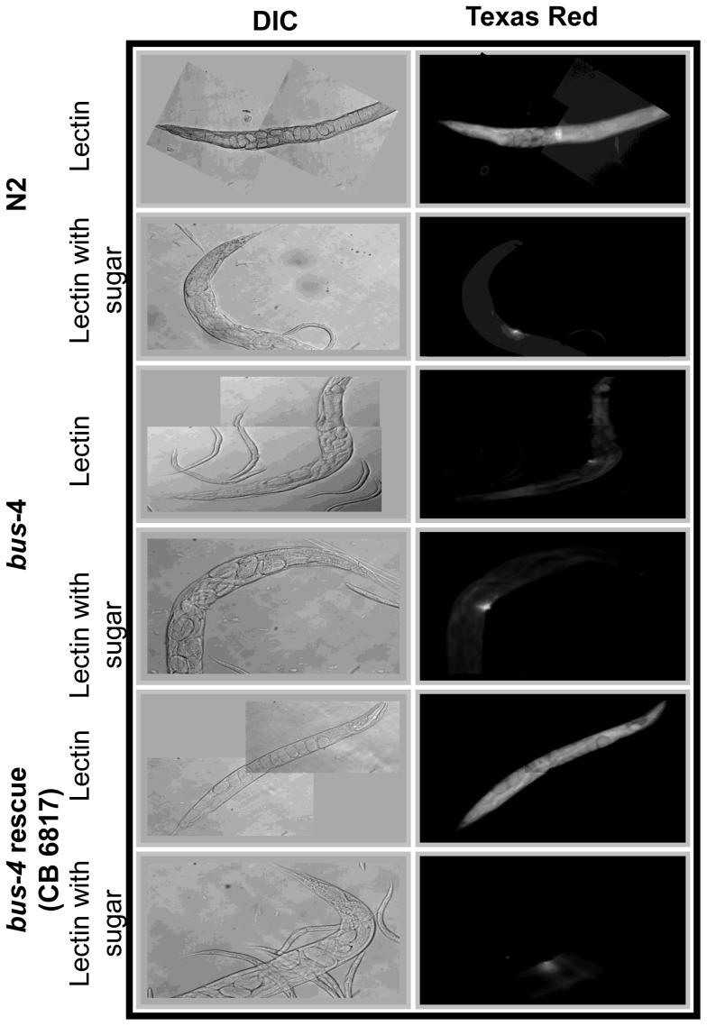
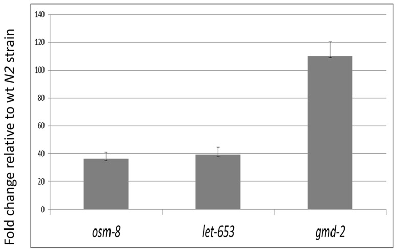
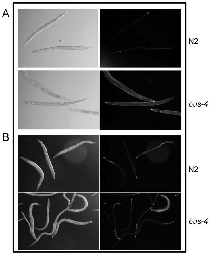
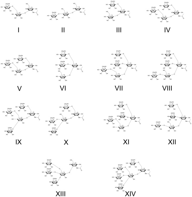
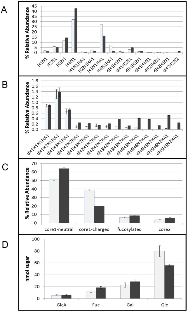
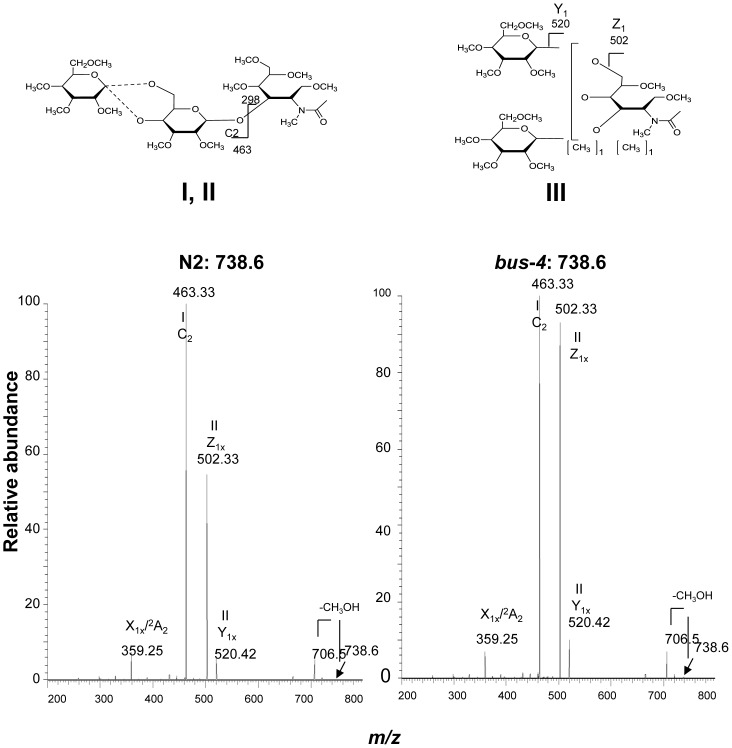
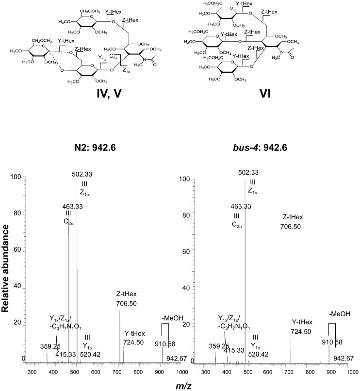
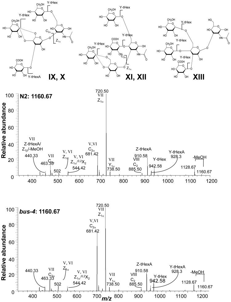
Caenorabditis elegans bus-4 glycosyltransferase mutants are resistant to infection by Microbacterium nematophilum, Yersinia pestis and Yersinia pseudotuberculosis and have altered susceptibility to two Leucobacter species Verde1 and Verde2. Our objective in this study was to define the glycosylation changes leading to this phenotype to better understand how these changes lead to pathogen resistance. We performed MALDI-TOF MS, tandem MS and GC/MS experiments to reveal fine structural detail for the bus-4 N- and O-glycan pools. We observed dramatic changes in O-glycans and moderate ones in N-glycan pools compared to the parent strain. Ce core-I glycans, the nematode's mucin glycan equivalent, were doubled in abundance, halved in charge and bore shifts in terminal substitutions. The fucosyl O-glycans, Ce core-II and neutral fucosyl forms, were also increased in abundance as were fucosyl N-glycans. Quantitative expression analysis revealed that two mucins, let-653 and osm-8, were upregulated nearly 40 fold and also revealed was a dramatic increase in GDP-Man 4,6 dehydratease expression. We performed detailed lectin binding studies that showed changes in glycoconjugates in the surface coat, cuticle surface and intestine. The combined changes in cell surface glycoconjugate distribution, increased abundance and altered properties of mucin provide an environment where likely the above pathogens are not exposed to normal glycoconjugate dependent cues leading to barriers to these bacterial infections.
秀丽隐杆线虫bus-4糖基转移酶突变体对嗜线虫微杆菌、鼠疫耶尔森菌和假结核耶尔森菌的感染具有抗性,并且对两种勒克氏菌属Verde1和Verde2的易感性发生了改变。我们在本研究中的目标是确定导致这种表型的糖基化变化,以便更好地理解这些变化如何导致病原体抗性。我们进行了基质辅助激光解吸电离飞行时间质谱(MALDI-TOF MS)、串联质谱和气相色谱/质谱(GC/MS)实验,以揭示bus-4 N-聚糖和O-聚糖库的精细结构细节。与亲本菌株相比,我们观察到O-聚糖有显著变化,而N-聚糖库有中度变化。线虫的粘蛋白聚糖等效物Ce核心I聚糖的丰度增加了一倍,电荷减半,末端取代发生了变化。岩藻糖基O-聚糖、Ce核心II和中性岩藻糖基形式的丰度也增加了,岩藻糖基N-聚糖也是如此。定量表达分析表明,两种粘蛋白let-65和osm-8的表达上调了近40倍,并且还发现GDP-甘露糖4,6脱水酶的表达显著增加。我们进行了详细的凝集素结合研究,结果显示表面被膜、角质层表面和肠道中的糖缀合物发生了变化。细胞表面糖缀合物分布的综合变化、粘蛋白丰度的增加和性质的改变提供了一个环境,在这个环境中,上述病原体可能无法接触到正常的依赖糖缀合物的信号,从而形成了对这些细菌感染的屏障。