Sun Yujing, Zhou Gengyin, Gui Ting, Shimokado Aiko, Nakanishi Masako, Oikawa Kosuke, Sato Fuyuki, Muragaki Yasuteru
1] First Department of Pathology, Wakayama Medical University School of Medicine, 811-1 Kimiidera, Wakayama 641-0012, Japan [2] Department of Pathology, School of Medicine, Shandong University, Jinan Wen Hua Xi Road 44, Jinan 250012, PR China.
Department of Pathology, School of Medicine, Shandong University, Jinan Wen Hua Xi Road 44, Jinan 250012, PR China.
Sci Rep. 2014 Oct 9;4:6563. doi: 10.1038/srep06563.
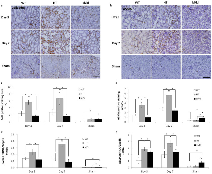
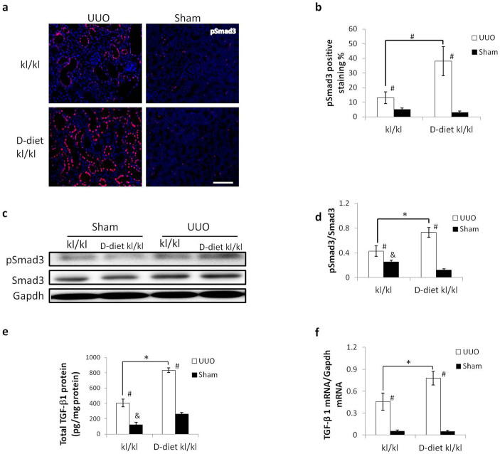
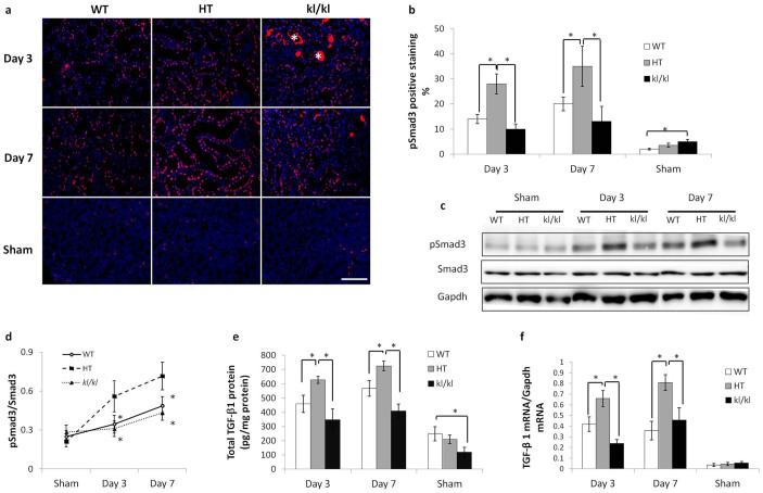
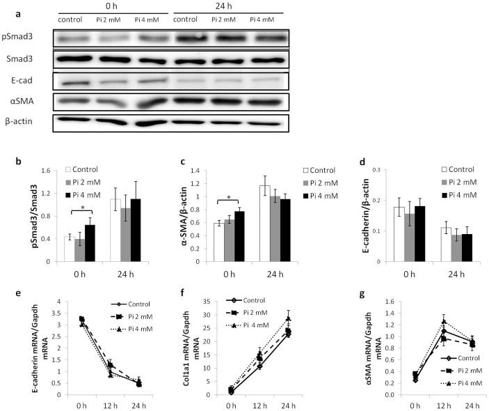
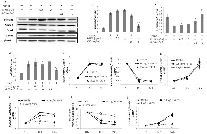
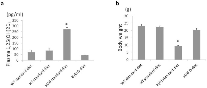
Previous studies have suggested that Klotho provides reno-protection against unilateral ureteral obstruction (UUO)-induced renal tubulointerstitial fibrosis (RTF). Because the existing studies are mainly performed using heterozygous Klotho mutant (HT) mice, we focused on the effect of UUO on homozygous Klotho mutant (kl/kl) mice. UUO kidneys from HT mice showed a significantly higher level of RTF and TGF-β/Smad3 signaling than wild-type (WT) mice, whereas both were greatly suppressed in kl/kl mice. Primary proximal tubular epithelial culture cells isolated from kl/kl mice showed no suppression in TGF-β1-induced epithelial mesenchymal transition (EMT) compared to those from HT mice. In the renal epithelial cell line NRK52E, a large amount of inorganic phosphate (Pi), FGF23, or calcitriol was added to the medium to mimic the in vivo homeostasis of kl/kl mice. Neither Pi nor FGF23 antagonized TGF-β1-induced EMT. In contrast, calcitriol ameliorated TGF-β1-induced EMT in a dose dependent manner. A vitamin D3-deficient diet normalized the serum 1,25 (OH)2 vitamin D3 level in kl/kl mice and enhanced UUO-induced RTF and TGF-β/Smad3 signaling. In conclusion, the alleviation of UUO-induced RTF in kl/kl mice was due to the TGF-β1 signaling suppression caused by an elevated serum 1, 25(OH)2 vitamin D3.
先前的研究表明,α-klotho蛋白可为单侧输尿管梗阻(UUO)诱导的肾小管间质纤维化(RTF)提供肾脏保护作用。由于现有研究主要使用杂合型α-klotho突变体(HT)小鼠进行,我们重点研究了UUO对纯合型α-klotho突变体(kl/kl)小鼠的影响。与野生型(WT)小鼠相比,HT小鼠的UUO肾脏显示出显著更高水平的RTF和TGF-β/Smad3信号传导,而在kl/kl小鼠中这两者均受到极大抑制。与从HT小鼠分离的原代近端肾小管上皮培养细胞相比,从kl/kl小鼠分离的细胞在TGF-β1诱导的上皮-间质转化(EMT)中未表现出抑制作用。在肾上皮细胞系NRK52E中,向培养基中添加大量无机磷酸盐(Pi)、成纤维细胞生长因子23(FGF23)或骨化三醇以模拟kl/kl小鼠的体内稳态。Pi和FGF23均未拮抗TGF-β1诱导的EMT。相反,骨化三醇以剂量依赖方式改善了TGF-β1诱导的EMT。缺乏维生素D3的饮食使kl/kl小鼠的血清1,25(OH)2维生素D3水平正常化,并增强了UUO诱导的RTF和TGF-β/Smad3信号传导。总之,kl/kl小鼠中UUO诱导的RTF减轻是由于血清1,25(OH)2维生素D3升高导致的TGF-β1信号传导抑制。