Cho Jang-Hee, Ryu Hye-Myung, Oh Eun-Joo, Yook Ju-Min, Ahn Ji-Sun, Jung Hee-Yeon, Choi Ji-Young, Park Sun-Hee, Kim Yong-Lim, Kwak Ihm Soo, Kim Chan-Duck
Department of Internal Medicine, Kyungpook National University School of Medicine, Daegu, Korea.
Department of Internal Medicine, Pusan National University Hospital, Busan, Korea.
PLoS One. 2016 Sep 8;11(9):e0162186. doi: 10.1371/journal.pone.0162186. eCollection 2016.
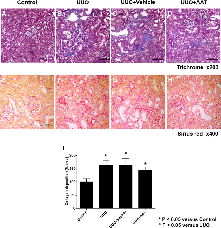
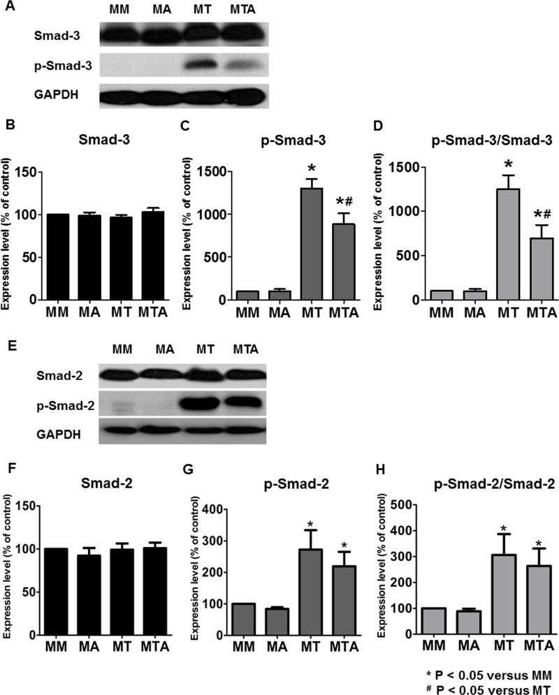
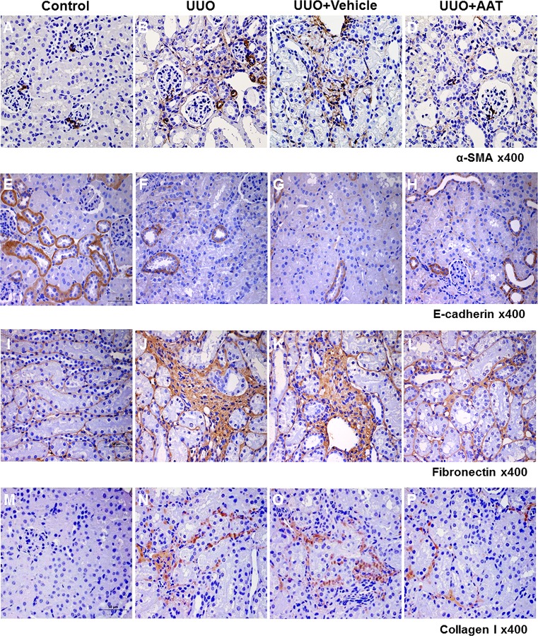
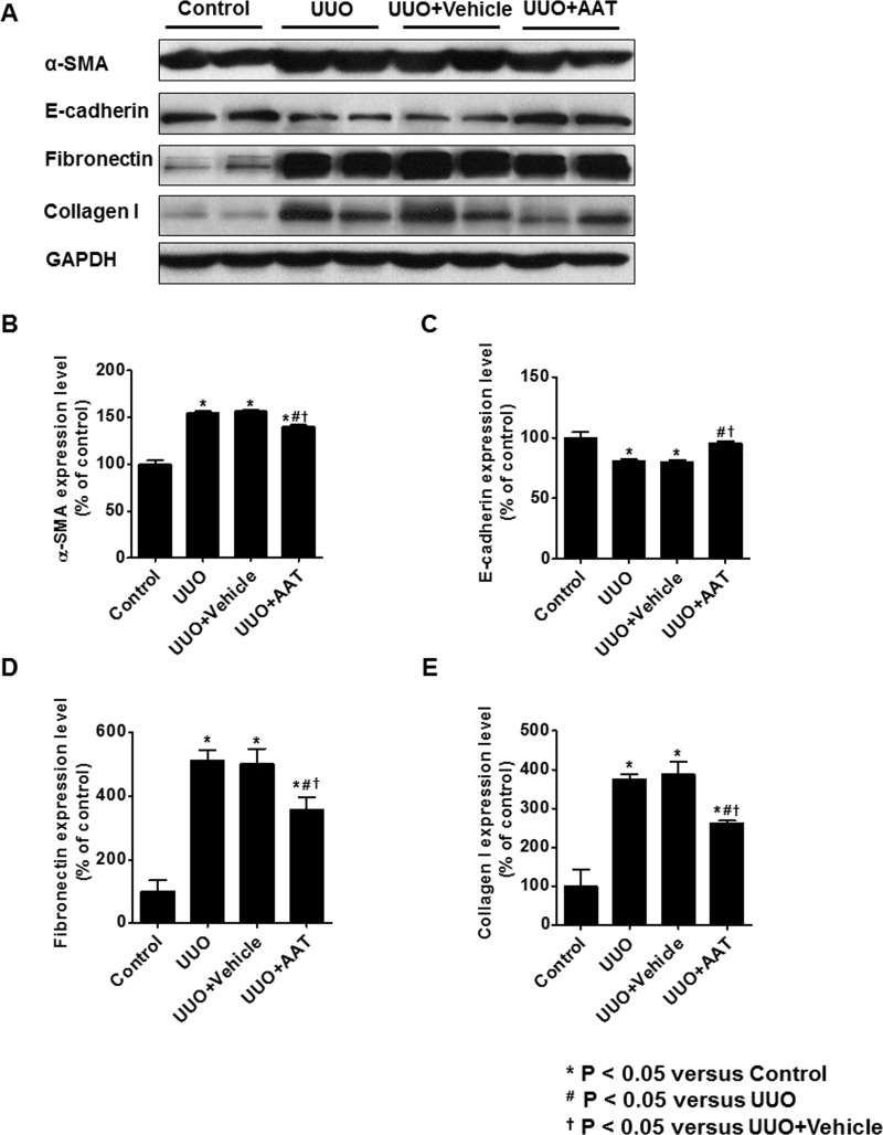
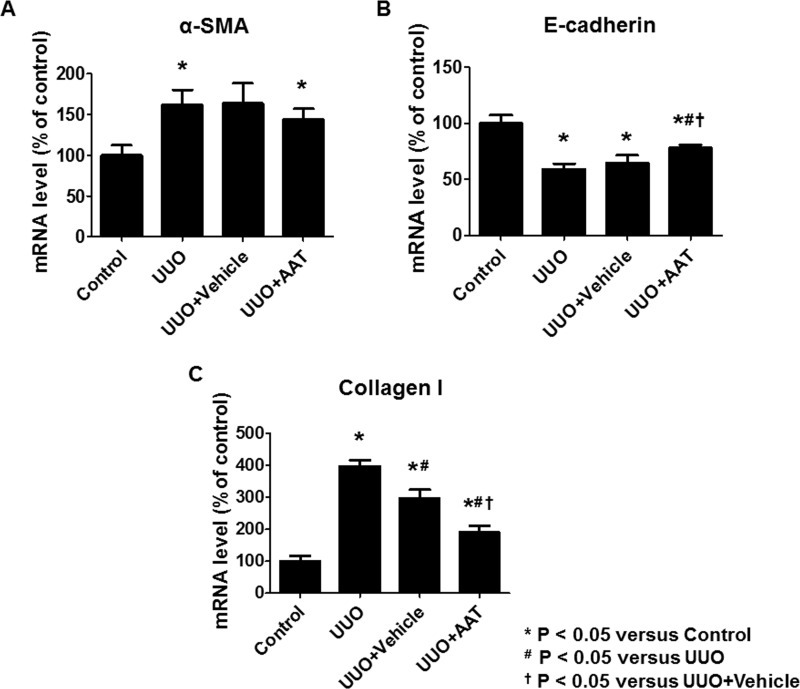
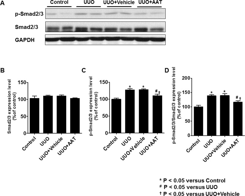
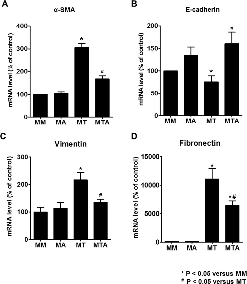
Alpha1-antitrypsin (AAT) exerts its anti-inflammatory effect through regulating the activity of serine proteinases. This study evaluated the inhibitory effects of AAT against the transforming growth factor (TGF)-β1 induced epithelial-to-mesenchymal transition (EMT) in unilateral ureter obstruction (UUO) mice and Madin-Darby canine kidney (MDCK) cells. C57BL/6 mice with induced UUO were injected intraperitoneally with AAT (80 mg/Kg) or vehicle for 7 days. MDCK cells were treated with TGF-β1 (2 ng/mL) for 48 hours to induce EMT, and co-treated with AAT (10 mg/mL) to inhibit the EMT. Masson's trichrome and Sirius red staining was used to estimate the extent of renal fibrosis in UUO mice. The expression of alpha-smooth muscle actin (α-SMA), vimentin, fibronectin, collagen I, and E-cadherin in MDCK cells and kidney tissue were evaluated. Masson's and Sirius red staining revealed that the area of renal fibrosis was significantly smaller in AAT treated UUO group compared with that of UUO and vehicle treated UUO groups. AAT treatment attenuated upregulation of Smad2/3 phosphorylation in UUO mouse model. Co-treatment of MDCK cells with TGF-β1 and AAT significantly attenuated the changes in the expression of α-SMA, vimentin, fibronectin, collagen I, and E-cadherin. AAT also decreased the phosphorylated Smad3 expression and the phosphorylated Smad3/Smad3 ratio in MDCK cells. AAT treatment inhibited EMT induced by TGF-β1 in MDCK cells and attenuated renal fibrosis in the UUO mouse model. The results of this work suggest that AAT could inhibit the process of EMT through the suppression of TGF-β/Smad3 signaling.
α1-抗胰蛋白酶(AAT)通过调节丝氨酸蛋白酶的活性发挥其抗炎作用。本研究评估了AAT对单侧输尿管梗阻(UUO)小鼠和麦迪逊-达比犬肾(MDCK)细胞中转化生长因子(TGF)-β1诱导的上皮-间质转化(EMT)的抑制作用。诱导UUO的C57BL/6小鼠腹腔注射AAT(80 mg/Kg)或溶剂,持续7天。MDCK细胞用TGF-β1(2 ng/mL)处理48小时以诱导EMT,并与AAT(10 mg/mL)共同处理以抑制EMT。采用Masson三色染色和天狼星红染色评估UUO小鼠肾纤维化程度。评估MDCK细胞和肾组织中α-平滑肌肌动蛋白(α-SMA)、波形蛋白、纤连蛋白、胶原蛋白I和E-钙黏蛋白的表达。Masson染色和天狼星红染色显示,与UUO组和溶剂处理的UUO组相比,AAT处理的UUO组肾纤维化面积显著更小。AAT处理减弱了UUO小鼠模型中Smad2/3磷酸化的上调。TGF-β1与AAT共同处理MDCK细胞显著减弱了α-SMA、波形蛋白、纤连蛋白、胶原蛋白I和E-钙黏蛋白表达的变化。AAT还降低了MDCK细胞中磷酸化Smad3的表达以及磷酸化Smad3/Smad3比值。AAT处理抑制了TGF-β1诱导的MDCK细胞中的EMT,并减轻了UUO小鼠模型中的肾纤维化。本研究结果表明,AAT可通过抑制TGF-β/Smad3信号传导来抑制EMT过程。