Velroyen Astrid, Bech Martin, Zanette Irene, Schwarz Jolanda, Rack Alexander, Tympner Christiane, Herrler Tanja, Staab-Weijnitz Claudia, Braunagel Margarita, Reiser Maximilian, Bamberg Fabian, Pfeiffer Franz, Notohamiprodjo Mike
Chair of Biomedical Physics, Department of Physics (E17), Munich, Bavaria, Germany.
Chair of Biomedical Physics, Department of Physics (E17), Munich, Bavaria, Germany; Medical Radiation Physics, Lund University, Lund, Sweden.
PLoS One. 2014 Oct 9;9(10):e109562. doi: 10.1371/journal.pone.0109562. eCollection 2014.
The aim of the study was to investigate microstructural changes occurring in unilateral renal ischemia-reperfusion injury in a murine animal model using synchrotron radiation.
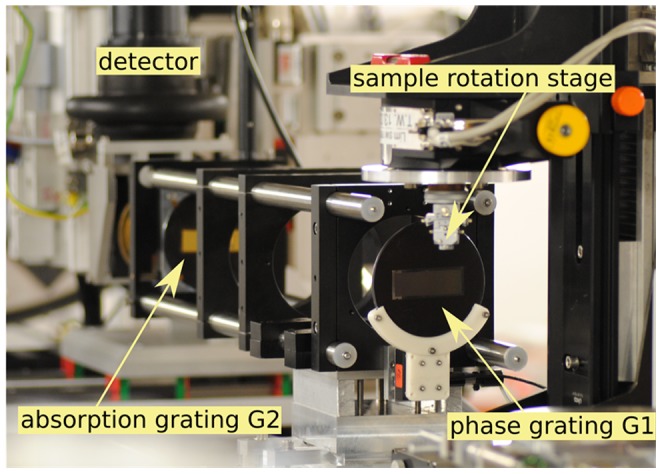
The effects of renal ischemia-reperfusion were investigated in a murine animal model of unilateral ischemia. Kidney samples were harvested on day 18. Grating-Based Phase-Contrast Imaging (GB-PCI) of the paraffin-embedded kidney samples was performed at a Synchrotron Radiation Facility (beam energy of 19 keV). To obtain phase information, a two-grating Talbot interferometer was used applying the phase stepping technique. The imaging system provided an effective pixel size of 7.5 µm. The resulting attenuation and differential phase projections were tomographically reconstructed using filtered back-projection. Semi-automated segmentation and volumetry and correlation to histopathology were performed.
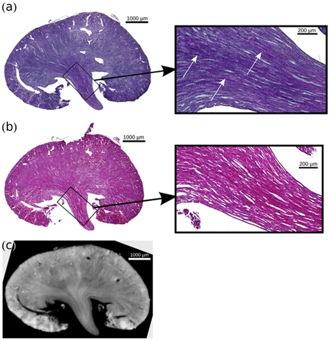
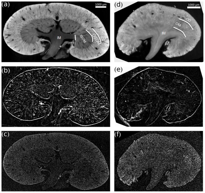
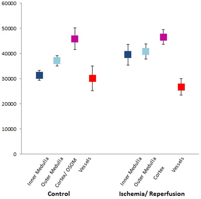
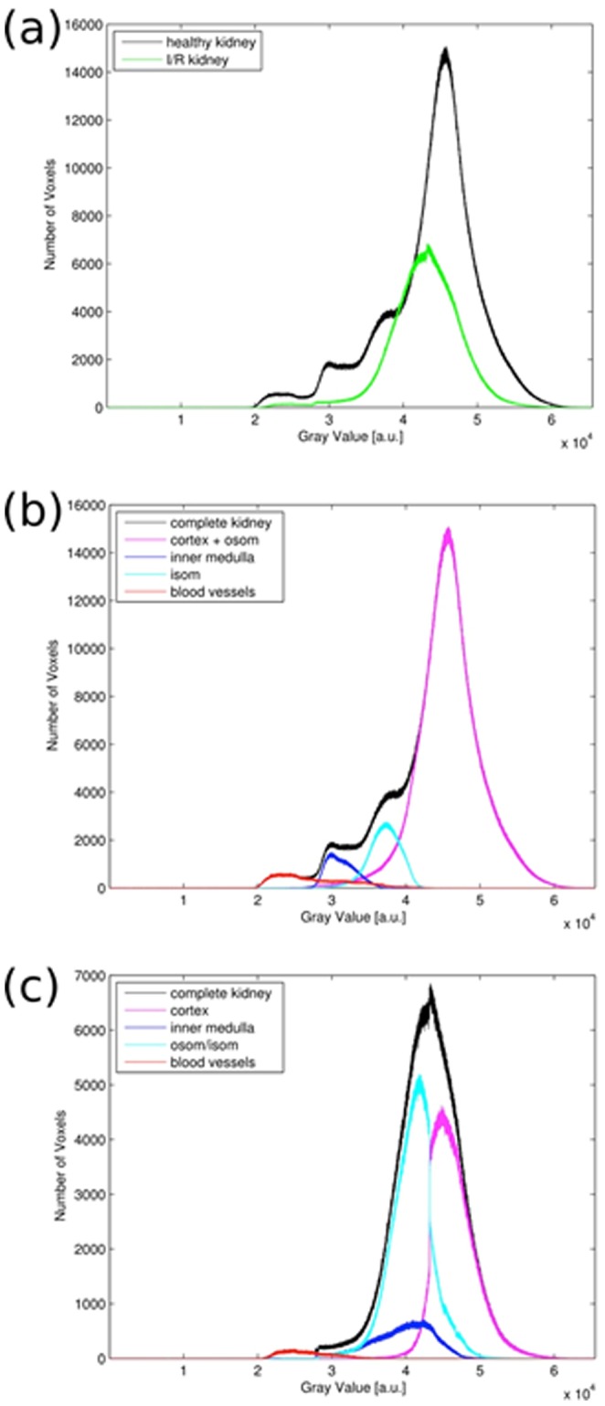
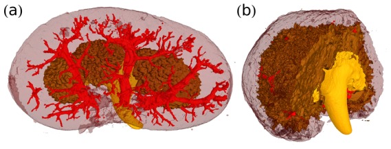
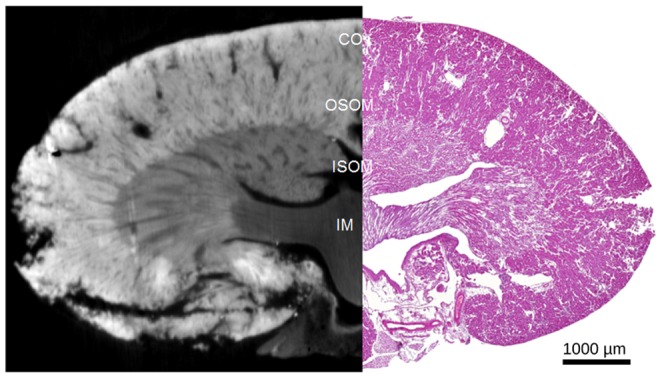
GB-PCI provided good discrimination of the cortex, outer and inner medulla in non-ischemic control kidneys. Post-ischemic kidneys showed a reduced compartmental differentiation, particularly of the outer stripe of the outer medulla, which could not be differentiated from the inner stripe. Compared to the contralateral kidney, after ischemia a volume loss was detected, while the inner medulla mainly retained its volume (ratio 0.94). Post-ischemic kidneys exhibited severe tissue damage as evidenced by tubular atrophy and dilatation, moderate inflammatory infiltration, loss of brush borders and tubular protein cylinders.
In conclusion GB-PCI with synchrotron radiation allows for non-destructive microstructural assessment of parenchymal kidney disease and vessel architecture. If translation to lab-based approaches generates sufficient density resolution, and with a time-optimized image analysis protocol, GB-PCI may ultimately serve as a non-invasive, non-enhanced alternative for imaging of pathological changes of the kidney.
本研究旨在利用同步辐射研究小鼠动物模型中单侧肾缺血再灌注损伤时发生的微观结构变化。
在单侧缺血的小鼠动物模型中研究肾缺血再灌注的影响。于第18天采集肾脏样本。在同步辐射设施(束流能量为19 keV)对石蜡包埋的肾脏样本进行基于光栅的相衬成像(GB-PCI)。为获取相位信息,使用双光栅Talbot干涉仪并应用相移技术。成像系统提供的有效像素尺寸为7.5 µm。使用滤波反投影对所得的衰减和微分相位投影进行断层重建。进行半自动分割、体积测量并与组织病理学进行相关性分析。
GB-PCI能很好地区分非缺血对照肾脏中的皮质、外髓和内髓。缺血后的肾脏显示出分区差异减小,尤其是外髓外带,无法与内带区分开来。与对侧肾脏相比,缺血后检测到体积减小,而内髓主要保持其体积(比例为0.94)。缺血后的肾脏表现出严重的组织损伤,表现为肾小管萎缩和扩张、中度炎症浸润、刷状缘丧失和肾小管蛋白管型。
总之,同步辐射GB-PCI可对肾实质疾病和血管结构进行非破坏性微观结构评估。如果转化为基于实验室的方法能产生足够的密度分辨率,并采用经过时间优化的图像分析方案,GB-PCI最终可能成为肾脏病理变化成像的一种非侵入性、无需增强的替代方法。