Radosevich Andrew J, Mutyal Nikhil N, Rogers Jeremy D, Gould Bradley, Hensing Thomas A, Ray Daniel, Backman Vadim, Roy Hemant K
Biomedical Engineering Department, Northwestern University, Evanston, Illinois, United States of America.
Biomedical Engineering Department, University of Wisconsin, Madison, Wisconsin, United States of America.
PLoS One. 2014 Oct 9;9(10):e110157. doi: 10.1371/journal.pone.0110157. eCollection 2014.
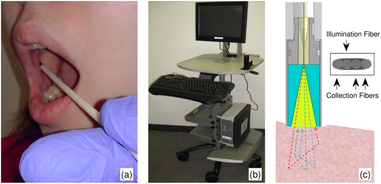
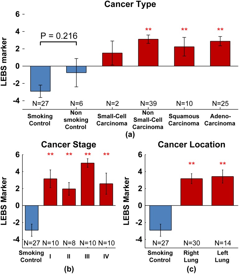
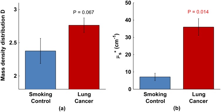
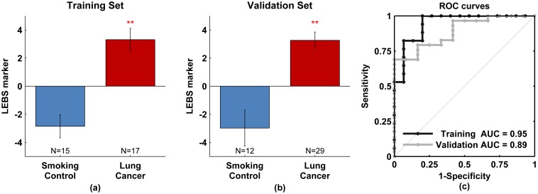
Lung cancer remains the leading cause of cancer deaths in the US with >150,000 deaths per year. In order to more effectively reduce lung cancer mortality, more sophisticated screening paradigms are needed. Previously, our group demonstrated the use of low-coherence enhanced backscattering (LEBS) spectroscopy to detect and quantify the micro/nano-architectural correlates of colorectal and pancreatic field carcinogenesis. In the lung, the buccal (cheek) mucosa has been suggested as an excellent surrogate site in the "field of injury". We, therefore, wanted to assess whether LEBS could similarly sense the presence of lung. To this end, we applied a fiber-optic LEBS probe to a dataset of 27 smokers without diagnosed lung cancer (controls) and 46 with lung cancer (cases), which was divided into a training and a blinded validation set (32 and 41 subjects, respectively). LEBS readings of the buccal mucosa were taken from the oral cavity applying gentle contact. The diagnostic LEBS marker was notably altered in patients harboring lung cancer compared to smoking controls. The prediction rule developed on training set data provided excellent diagnostics with 94% sensitivity, 80% specificity, and 95% accuracy. Applying the same threshold to the blinded validation set yielded 79% sensitivity and 83% specificity. These results were not confounded by patient demographics or impacted by cancer type or location. Moreover, the prediction rule was robust across all stages of cancer including stage I. We envision the use of LEBS as the first part of a two-step paradigm shift in lung cancer screening in which patients with high LEBS risk markers are funnelled into more invasive screening for confirmation.
肺癌仍是美国癌症死亡的主要原因,每年有超过15万人死亡。为了更有效地降低肺癌死亡率,需要更精密的筛查模式。此前,我们团队展示了利用低相干增强背向散射(LEBS)光谱技术来检测和量化结直肠癌和胰腺癌场癌发生的微观/纳米结构关联。在肺部,颊(脸颊)黏膜被认为是“损伤区域”中的一个极佳替代部位。因此,我们想评估LEBS是否同样能检测出肺部病变。为此,我们将光纤LEBS探头应用于27名未被诊断出患有肺癌的吸烟者(对照组)和46名患有肺癌的患者(病例组)的数据集,该数据集被分为训练集和盲法验证集(分别为32名和41名受试者)。通过轻轻接触口腔来获取颊黏膜的LEBS读数。与吸烟对照组相比,患有肺癌的患者的诊断性LEBS标志物有显著变化。基于训练集数据开发的预测规则具有出色的诊断能力,灵敏度为94%,特异性为80%,准确率为95%。将相同阈值应用于盲法验证集,灵敏度为79%,特异性为83%。这些结果不受患者人口统计学因素的干扰,也不受癌症类型或位置的影响。此外,该预测规则在包括I期在内的所有癌症阶段都很稳健。我们设想将LEBS用作肺癌筛查两步范式转变的第一步,即把具有高LEBS风险标志物的患者引导至更具侵入性的筛查以进行确诊。