Department of Biomedical Engineering, Erasmus MC, P.O. Box 2040, 3000 CA Rotterdam, The Netherlands ; Interuniversity Cardiology Institute of The Netherlands - Netherlands Heart Institute, P.O. Box 19258, 3501 DG Utrecht, The Netherlands.
Department of Biomedical Engineering, Erasmus MC, P.O. Box 2040, 3000 CA Rotterdam, The Netherlands.
Photoacoustics. 2013 Dec 5;2(1):12-20. doi: 10.1016/j.pacs.2013.11.003. eCollection 2014 Mar.
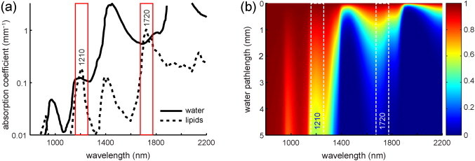
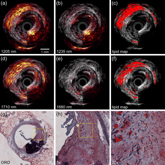
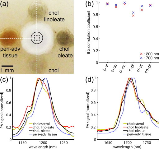
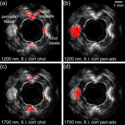
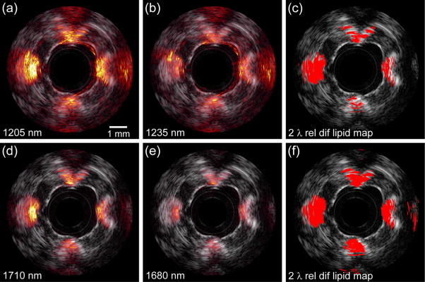
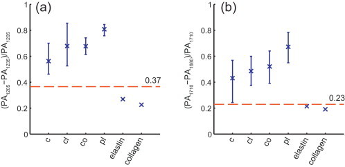
Spectroscopic intravascular photoacoustic imaging (sIVPA) has shown promise to detect and distinguish lipids in atherosclerotic plaques. sIVPA generally utilizes one of the two high absorption bands in the lipid absorption spectrum at 1.2 μm and 1.7 μm. Specific absorption signatures of various lipid compounds within the bands in either wavelength range can potentially be used to differentiate between plaque lipids and peri-adventitial lipids. With the aim to quantify any differences between the two bands, we performed combined sIVPA imaging in both absorption bands on a vessel phantom and an atherosclerotic human coronary artery ex vivo. Lipid detection in a human atherosclerotic lesion with sIVPA required lower pulse energy at 1.7 μm than at 1.2 μm (0.4 mJ versus 1.2 mJ). The imaging depth was twice as large at 1.2 μm compared to 1.7 μm. Adequate differentiation between plaque and peri-adventitial lipids was achieved at 1.2 μm only.
光谱学血管内光声成像(sIVPA)已显示出在检测和区分动脉粥样硬化斑块中的脂质方面的潜力。sIVPA 通常利用脂质吸收光谱在 1.2μm 和 1.7μm 处的两个高吸收带之一。在任一波长范围内,带内各种脂质化合物的特定吸收特征可用于区分斑块脂质和血管外膜脂质。为了量化两个波段之间的任何差异,我们在血管模型和离体动脉粥样硬化人冠状动脉上进行了两个吸收波段的联合 sIVPA 成像。在 sIVPA 中检测人类动脉粥样硬化病变中的脂质需要在 1.7μm 处的脉冲能量低于 1.2μm(0.4mJ 对 1.2mJ)。1.2μm 的成像深度是 1.7μm 的两倍。仅在 1.2μm 处可实现斑块和血管外膜脂质的充分区分。