Margheri Francesca, Papucci Laura, Schiavone Nicola, D'Agostino Riccardo, Trigari Silvana, Serratì Simona, Laurenzana Anna, Biagioni Alessio, Luciani Cristina, Chillà Anastasia, Andreucci Elena, Del Rosso Tommaso, Margheri Giancarlo, Del Rosso Mario, Fibbi Gabriella
Department of Experimental and Clinical Biomedical Sciences, Section of Experimental Pathology and Oncology, University of Florence, Florence, Italy.
J Cell Mol Med. 2015 Jan;19(1):113-23. doi: 10.1111/jcmm.12410. Epub 2014 Oct 14.
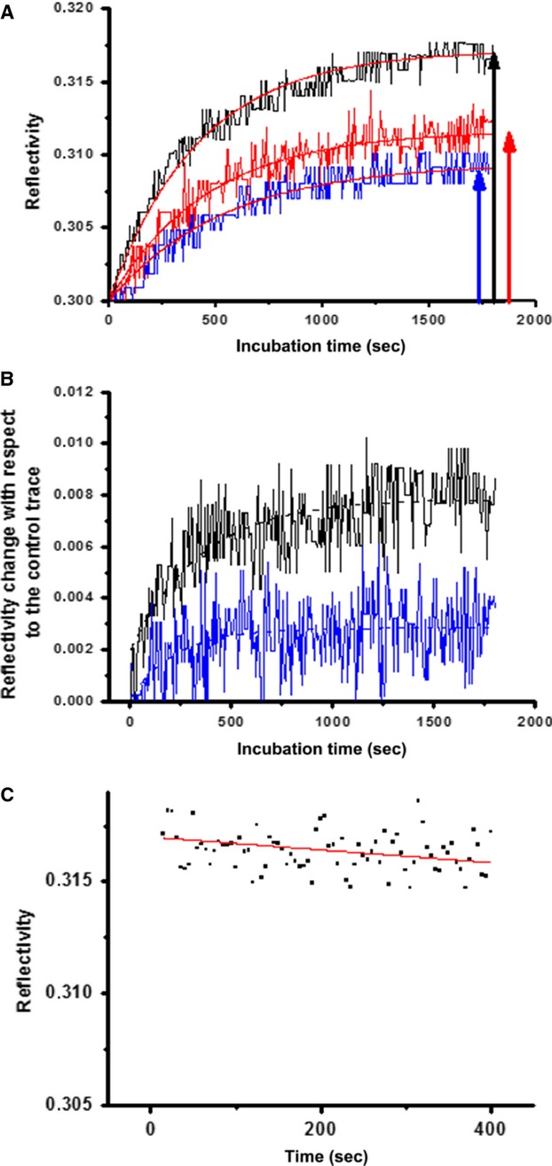
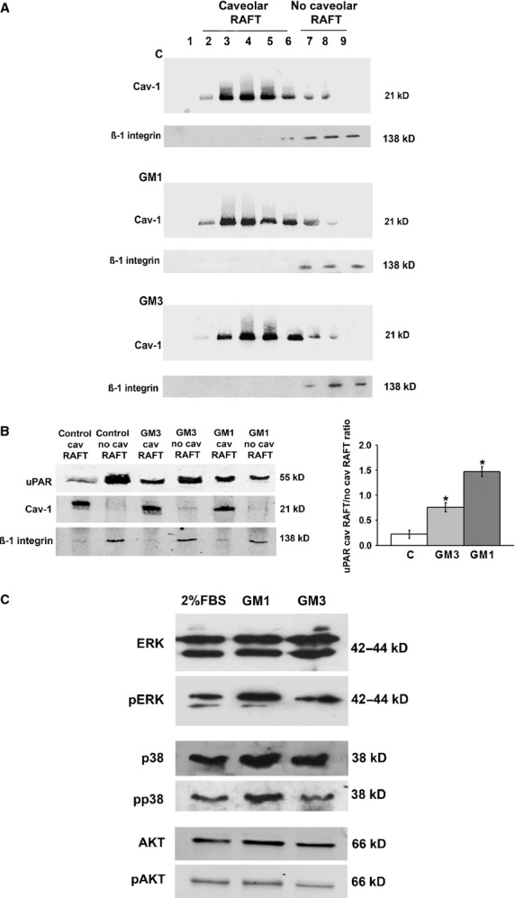
Gangliosides and the urokinase plasminogen activator receptor (uPAR) tipically partition in specialized membrane microdomains called lipid-rafts. uPAR becomes functionally important in fostering angiogenesis in endothelial progenitor cells (EPCs) upon recruitment in caveolar-lipid rafts. Moreover, cell membrane enrichment with exogenous GM1 ganglioside is pro-angiogenic and opposite to the activity of GM3 ganglioside. On these basis, we first checked the interaction of uPAR with membrane models enriched with GM1 or GM3, relying on the adoption of solid-supported mobile bilayer lipid membranes with raft-like composition formed onto solid hydrophilic surfaces, and evaluated by surface plasmon resonance (SPR) the extent of uPAR recruitment. We estimated the apparent dissociation constants of uPAR-GM1/GM3 complexes. These preliminary observations, indicating that uPAR binds preferentially to GM1-enriched biomimetic membranes, were validated by identifying a pro-angiogenic activity of GM1-enriched EPCs, based on GM1-dependent uPAR recruitment in caveolar rafts. We have observed that addition of GM1 to EPCs culture medium promotes matrigel invasion and capillary morphogenesis, as opposed to the anti-angiogenesis activity of GM3. Moreover, GM1 also stimulates MAPKinases signalling pathways, typically associated with an angiogenesis program. Caveolar-raft isolation and Western blotting of uPAR showed that GM1 promotes caveolar-raft partitioning of uPAR, as opposed to control and GM3-challenged EPCs. By confocal microscopy, we have shown that in EPCs uPAR is present on the surface in at least three compartments, respectively, associated to GM1, GM3 and caveolar rafts. Following GM1 exogenous addition, the GM3 compartment is depleted of uPAR which is recruited within caveolar rafts thereby triggering angiogenesis.
神经节苷脂和尿激酶型纤溶酶原激活物受体(uPAR)通常分布于称为脂筏的特殊膜微区中。在内皮祖细胞(EPCs)中,uPAR募集到小窝脂筏后,在促进血管生成方面发挥重要功能。此外,用外源性GM1神经节苷脂富集细胞膜具有促血管生成作用,这与GM3神经节苷脂的活性相反。基于这些,我们首先检测了uPAR与富含GM1或GM3的膜模型之间的相互作用,采用在固体亲水表面形成的具有类筏状组成的固体支持移动双层脂质膜,并通过表面等离子体共振(SPR)评估uPAR募集的程度。我们估算了uPAR - GM1/GM3复合物的表观解离常数。这些初步观察结果表明uPAR优先结合富含GM1的仿生膜,通过鉴定富含GM1的EPCs的促血管生成活性得到了验证,该活性基于小窝脂筏中GM1依赖的uPAR募集。我们观察到,向EPCs培养基中添加GM1可促进基质胶侵袭和毛细血管形态发生,这与GM3的抗血管生成活性相反。此外,GM1还刺激丝裂原活化蛋白激酶信号通路,该通路通常与血管生成程序相关。小窝脂筏分离及uPAR的蛋白质印迹分析表明,与对照和GM3处理的EPCs相反,GM1促进uPAR在小窝脂筏中的分布。通过共聚焦显微镜,我们发现EPCs表面的uPAR至少存在于三个区室中,分别与GM1、GM3和小窝脂筏相关。外源性添加GM1后,GM3区室中的uPAR减少,其被募集到小窝脂筏中,从而触发血管生成。