Department of Medical Imaging, the Second Hospital of Hebei Medical University, Shijiazhuang, Hebei Province, China.
Department of Endocrinology, the Second Hospital of Hebei Medical University, Shijiazhuang, Hebei Province, China.
Neural Regen Res. 2014 Aug 15;9(16):1548-56. doi: 10.4103/1673-5374.139482.
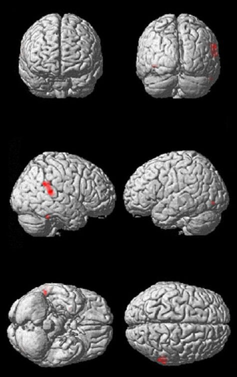
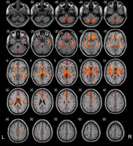
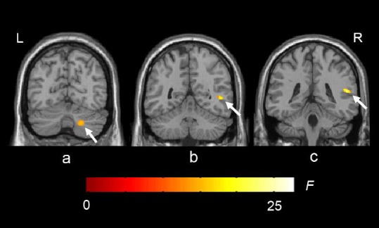
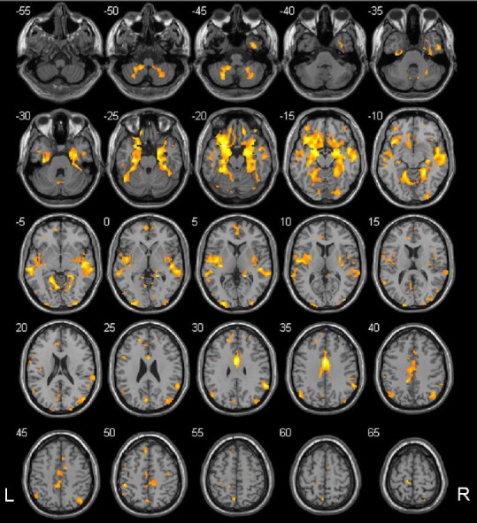
Voxel-based morphometry has been used in the study of alterations in brain structure in type 1 diabetes mellitus patients. These changes are associated with clinical indices. The age at onset, pathogenesis, and treatment of type 1 diabetes mellitus are different from those for type 2 diabetes mellitus. Thus, type 1 and type 2 diabetes mellitus may have different impacts on brain structure. Only a few studies of the alterations in brain structure in type 2 diabetes mellitus patients using voxel-based morphometry have been conducted, with inconsistent results. We detected subtle changes in the brain structure of 23 cases of type 2 diabetes mellitus, and demonstrated that there was no significant difference between the total volume of gray and white matter of the brain of type 2 diabetes mellitus patients and that in controls. Regional atrophy of gray matter mainly occurred in the right temporal and left occipital cortex, while regional atrophy of white matter involved the right temporal lobe and the right cerebellar hemisphere. The ankle-brachial index in patients with type 2 diabetes mellitus strongly correlated with the volume of brain regions in the default mode network. The ankle-brachial index, followed by the level of glycosylated hemoglobin, most strongly correlated with the volume of gray matter in the right temporal lobe. These data suggest that voxel-based morphometry could detect small structural changes in patients with type 2 diabetes mellitus. Early macrovascular atherosclerosis may play a crucial role in subtle brain atrophy in type 2 diabetes mellitus patients, with chronic hyperglycemia playing a lesser role.
体素形态计量学已被用于研究 1 型糖尿病患者大脑结构的变化。这些变化与临床指标有关。1 型糖尿病的发病年龄、发病机制和治疗方法与 2 型糖尿病不同。因此,1 型和 2 型糖尿病可能对大脑结构有不同的影响。只有少数使用体素形态计量学研究 2 型糖尿病患者大脑结构变化的研究,结果不一致。我们检测了 23 例 2 型糖尿病患者的大脑结构细微变化,结果表明 2 型糖尿病患者大脑灰质和白质的总体积与对照组无显著差异。灰质的区域性萎缩主要发生在右侧颞叶和左侧枕叶,而白质的区域性萎缩则涉及右侧颞叶和右侧小脑半球。2 型糖尿病患者的踝臂指数与默认模式网络中大脑区域的体积呈强烈相关性。踝臂指数,其次是糖化血红蛋白水平,与右侧颞叶灰质体积的相关性最强。这些数据表明,体素形态计量学可以检测到 2 型糖尿病患者的微小结构变化。早期大血管粥样硬化可能在 2 型糖尿病患者的细微脑萎缩中起关键作用,而慢性高血糖的作用较小。