Moran Chris, Phan Thanh G, Chen Jian, Blizzard Leigh, Beare Richard, Venn Alison, Münch Gerald, Wood Amanda G, Forbes Josephine, Greenaway Timothy M, Pearson Susan, Srikanth Velandai
Corresponding author: Velandai Srikanth,
Diabetes Care. 2013 Dec;36(12):4036-42. doi: 10.2337/dc13-0143. Epub 2013 Aug 12.
Type 2 diabetes (T2DM) is associated with brain atrophy and cerebrovascular disease. We aimed to define the regional distribution of brain atrophy in T2DM and to examine whether atrophy or cerebrovascular lesions are feasible links between T2DM and cognitive function.
This cross-sectional study used magnetic resonance imaging (MRI) scans and cognitive tests in 350 participants with T2DM and 363 participants without T2DM. With voxel-based morphometry, we studied the regional distribution of atrophy in T2DM. We measured cerebrovascular lesions (infarcts, microbleeds, and white matter hyperintensity [WMH] volume) and atrophy (gray matter, white matter, and hippocampal volumes) while blinded to T2DM status. With use of multivariable regression, we examined for mediation or effect modification of the association between T2DM and cognitive measures by MRI measures.
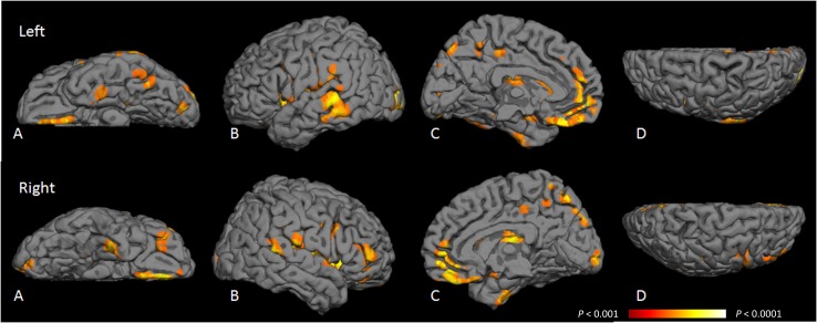
T2DM was associated with more cerebral infarcts and lower total gray, white, and hippocampal volumes (all P < 0.05) but not with microbleeds or WMH. T2DM-related gray matter loss was distributed mainly in medial temporal, anterior cingulate, and medial frontal lobes, and white matter loss was distributed in frontal and temporal regions. T2DM was associated with poorer visuospatial construction, planning, visual memory, and speed (P ≤ 0.05) independent of age, sex, education, and vascular risk factors. The strength of these associations was attenuated by almost one-half when adjusted for hippocampal and total gray volumes but was unchanged by adjustment for cerebrovascular lesions or white matter volume.
Cortical atrophy in T2DM resembles patterns seen in preclinical Alzheimer disease. Neurodegeneration rather than cerebrovascular lesions may play a key role in T2DM-related cognitive impairment.
2型糖尿病(T2DM)与脑萎缩和脑血管疾病相关。我们旨在确定T2DM患者脑萎缩的区域分布,并研究萎缩或脑血管病变是否是T2DM与认知功能之间可能的联系。
这项横断面研究对350名T2DM患者和363名非T2DM患者进行了磁共振成像(MRI)扫描和认知测试。采用基于体素的形态学测量方法,我们研究了T2DM患者萎缩的区域分布。在对T2DM状态不知情的情况下,我们测量了脑血管病变(梗死灶、微出血和白质高信号[WMH]体积)和萎缩(灰质、白质和海马体体积)。通过多变量回归分析,我们研究了MRI测量指标对T2DM与认知指标之间关联的中介作用或效应修正作用。
T2DM与更多的脑梗死以及更低的总灰质、白质和海马体体积相关(所有P<0.05),但与微出血或WMH无关。T2DM相关的灰质损失主要分布在内侧颞叶、前扣带回和内侧额叶,白质损失分布在额叶和颞叶区域。独立于年龄、性别、教育程度和血管危险因素,T2DM与较差的视觉空间构建、计划、视觉记忆和速度相关(P≤0.05)。在调整海马体和总灰质体积后,这些关联的强度减弱了近一半,但在调整脑血管病变或白质体积后没有变化。
T2DM患者的皮质萎缩类似于临床前阿尔茨海默病的模式。神经退行性变而非脑血管病变可能在T2DM相关的认知障碍中起关键作用。