• 文献检索
  • 文档翻译
  • 深度研究
  • 学术资讯
  • Suppr Zotero 插件Zotero 插件
  • 邀请有礼
  • 套餐&价格
  • 历史记录
应用&插件
Suppr Zotero 插件Zotero 插件浏览器插件Mac 客户端Windows 客户端微信小程序
定价
高级版会员购买积分包购买API积分包
服务
文献检索文档翻译深度研究API 文档MCP 服务
关于我们
关于 Suppr公司介绍联系我们用户协议隐私条款
关注我们

Suppr 超能文献

核心技术专利:CN118964589B侵权必究
粤ICP备2023148730 号-1Suppr @ 2026

文献检索

告别复杂PubMed语法,用中文像聊天一样搜索,搜遍4000万医学文献。AI智能推荐,让科研检索更轻松。

立即免费搜索

文件翻译

保留排版,准确专业,支持PDF/Word/PPT等文件格式,支持 12+语言互译。

免费翻译文档

深度研究

AI帮你快速写综述,25分钟生成高质量综述,智能提取关键信息,辅助科研写作。

立即免费体验

一名75岁患有肺动脉高压的女性,存在部分肺静脉异常连接至上腔静脉,该连接跨越完整的房间隔并具有双心房连接。

Partial anomalous pulmonary venous connection to superior vena cava that overrides across the intact atrial septum and has bi-atrial connection in a 75-year-old female presenting with pulmonary hypertension.

作者信息

Wang Hong, Guan Hanxiong, Wang Dao Wen

机构信息

Division of Cardiology and Department of Internal Medicine, Tongji Hospital, Tongji Medical College of Huazhong University of Science and Technology, 1905 Jiefang Dadao, Wuhan 430030, PR China.

出版信息

BMC Cardiovasc Disord. 2014 Oct 25;14:149. doi: 10.1186/1471-2261-14-149.

DOI:10.1186/1471-2261-14-149
PMID:25344786
原文链接:https://pmc.ncbi.nlm.nih.gov/articles/PMC4219022/
Abstract

BACKGROUND

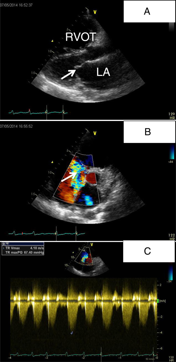
Partial anomalous venous connection (PAPVC) is a rare congenital heart disease where the blood flow from one or more pulmonary veins (but not all) returns to the right atrium or systemic venous circulation and is often associated with a sinus venosus atrial defect (SVD). Transthoracic echocardiography (TTE) can provide limited information for this anomaly and the diagnosis of this congenital defect has been a clinical challenge.

CASE PRESENTATION

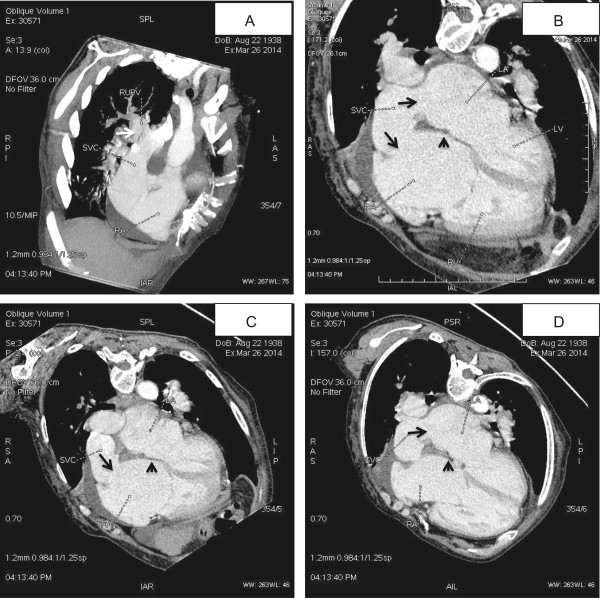
We report here a case of a 75-year-old female with adult-onset pulmonary arterial hypertension (PAH), hypoxemia and right-sided chamber dilatation. The diagnosis of PAPVC was made incidentally by multidetector computed tomographic angiography (MCTA) that was performed to exclude pulmonary embolism. In this type of PAPVC, the atrial septum is intact, the right upper pulmonary vein (RUPV) connects to the superior vena cava (SVC), and the SVC overrides across the atrial septum and has bi-atrial connection, all of which are clearly manifested by MCTA.

CONCLUSIONS

This case indicates the need to exclude a PAPVC and SVD in unexplained pulmonary hypertension, and MCTA is a reliable non-invasive imaging technique with high resolution and wide anatomic coverage. The case also demonstrates that the coexisting SVD with PAPVC is an anomalous venous connection instead of atrial septal defect (ASD) and its key feature is the overriding of SVC or IVC across the intact atrial septum.

摘要

背景

部分性肺静脉异位连接(PAPVC)是一种罕见的先天性心脏病,其中一条或多条(而非全部)肺静脉的血流回流至右心房或体静脉循环,且常与静脉窦型房间隔缺损(SVD)相关。经胸超声心动图(TTE)对这种异常情况提供的信息有限,而诊断这种先天性缺陷一直是一项临床挑战。

病例报告

我们在此报告一例75岁女性患者,患有成人起病的肺动脉高压(PAH)、低氧血症和右心腔扩张。PAPVC的诊断是在进行多排螺旋计算机断层血管造影(MCTA)以排除肺栓塞时偶然发现的。在这种类型的PAPVC中,房间隔完整,右上肺静脉(RUPV)连接至上腔静脉(SVC),且SVC跨越房间隔并具有双心房连接,所有这些在MCTA上均清晰显示。

结论

该病例表明,在不明原因的肺动脉高压中需要排除PAPVC和SVD,且MCTA是一种可靠的非侵入性成像技术,具有高分辨率和广泛的解剖覆盖范围。该病例还表明,PAPVC合并存在的SVD是一种肺静脉异位连接而非房间隔缺损(ASD),其关键特征是SVC或下腔静脉(IVC)跨越完整的房间隔。

https://cdn.ncbi.nlm.nih.gov/pmc/blobs/39b9/4219022/0c92c421ebc1/12872_2014_792_Fig2_HTML.jpg
https://cdn.ncbi.nlm.nih.gov/pmc/blobs/39b9/4219022/06fee48c1aa1/12872_2014_792_Fig1_HTML.jpg
https://cdn.ncbi.nlm.nih.gov/pmc/blobs/39b9/4219022/0c92c421ebc1/12872_2014_792_Fig2_HTML.jpg
https://cdn.ncbi.nlm.nih.gov/pmc/blobs/39b9/4219022/06fee48c1aa1/12872_2014_792_Fig1_HTML.jpg
https://cdn.ncbi.nlm.nih.gov/pmc/blobs/39b9/4219022/0c92c421ebc1/12872_2014_792_Fig2_HTML.jpg

相似文献

1
Partial anomalous pulmonary venous connection to superior vena cava that overrides across the intact atrial septum and has bi-atrial connection in a 75-year-old female presenting with pulmonary hypertension.一名75岁患有肺动脉高压的女性,存在部分肺静脉异常连接至上腔静脉,该连接跨越完整的房间隔并具有双心房连接。
BMC Cardiovasc Disord. 2014 Oct 25;14:149. doi: 10.1186/1471-2261-14-149.
2
Partial anomalous pulmonary venous connection: diagnosis by transesophageal echocardiography.部分性肺静脉异位连接:经食管超声心动图诊断
J Am Coll Cardiol. 1997 May;29(6):1351-8. doi: 10.1016/s0735-1097(97)82758-1.
3
Partial anomalous pulmonary venous connection to the right side of the heart.部分肺静脉异常连接至心脏右侧。
J Thorac Cardiovasc Surg. 1989 Nov;98(5 Pt 2):861-8.
4
A case of right superior vena cava draining into the left atrial roof.一例右上腔静脉引流至左心房顶部的病例。
Asian Cardiovasc Thorac Ann. 2016 Feb;24(2):158-61. doi: 10.1177/0218492314547544. Epub 2014 Aug 13.
5
Cardiac MRI and pulmonary MR angiography of sinus venosus defect and partial anomalous pulmonary venous connection in cause of right undiagnosed ventricular enlargement.心脏磁共振成像及肺磁共振血管造影在诊断不明的右心室增大病因中的应用:静脉窦缺损及部分性肺静脉异位连接
AJR Am J Roentgenol. 2009 Jan;192(1):259-66. doi: 10.2214/AJR.07.3430.
6
Repair of Partial Anomalous Pulmonary Venous Connection: A Rare Case Evaluated by Multi-Slice Computed Tomographic Angiography.部分性肺静脉异位连接的修复:一例通过多层计算机断层血管造影评估的罕见病例
Heart Surg Forum. 2018 Mar 8;21(2):E099-E100. doi: 10.1532/hsf.1871.
7
Sinus venosus syndrome: atrial septal defect or anomalous venous connection? A multiplane transoesophageal approach.静脉窦综合征:房间隔缺损还是异常静脉连接?多平面经食管途径。
Heart. 2002 Dec;88(6):634-8. doi: 10.1136/heart.88.6.634.
8
[Superior vena cava-right atrial appendage direct anastomosis for repair of partial anomalous pulmonary venous connection to superior vena cava].[上腔静脉-右心耳直接吻合术修复部分性肺静脉异位连接至上腔静脉]
Kyobu Geka. 1994 Apr;47(4):307-10.
9
Clinical Significance of Partial Anomalous Pulmonary Venous Connections (Isolated and Atrial Septal Defect Associated) Determined by Cardiovascular Magnetic Resonance.心血管磁共振对部分肺静脉异常连接(孤立性和房间隔缺损相关)的临床意义。
Circ Cardiovasc Imaging. 2021 Aug;14(8):e012371. doi: 10.1161/CIRCIMAGING.120.012371. Epub 2021 Aug 13.
10
Novel technique of repairing right partial anomalous pulmonary venous connection with intact atrial septum using in situ interatrial septum as a flap in a 68-year-old-woman: a case report.应用原位房间隔作为瓣片修复 68 岁女性完整房间隔右侧部分肺静脉连接异常的新方法:病例报告。
J Cardiothorac Surg. 2020 Sep 25;15(1):269. doi: 10.1186/s13019-020-01313-w.

引用本文的文献

1
Anatomical Insights Into Profound Pulmonary Venous Malformation in an Elderly Individual: A Report of a Rare Cadaveric Case.老年个体严重肺静脉畸形的解剖学见解:一例罕见尸体病例报告
Cureus. 2025 Jul 14;17(7):e87905. doi: 10.7759/cureus.87905. eCollection 2025 Jul.
2
A Rare Case of Partial Anomalous Pulmonary Venous Connection.部分性肺静脉异位连接1例罕见病例
Cureus. 2023 Dec 21;15(12):e50913. doi: 10.7759/cureus.50913. eCollection 2023 Dec.
3
Multimodality diagnostic imaging for anomalous pulmonary venous connections: a pictorial essay.

本文引用的文献

1
Right upper lobe partial anomalous pulmonary venous connection.右上叶部分性肺静脉异位连接
Case Rep Vasc Med. 2014;2014:249896. doi: 10.1155/2014/249896. Epub 2014 Feb 16.
2
Partial anomalous pulmonary venous return and atrial septal defect in adult patients detected with 128-slice multidetector computed tomography.128层多排螺旋计算机断层扫描检测成人患者的部分性肺静脉异位引流和房间隔缺损
J Cardiothorac Surg. 2011 Sep 30;6:126. doi: 10.1186/1749-8090-6-126.
3
Sinus venosus atrial septal defect in a 31-year-old female patient: a case for surgical repair.
多模态诊断成像在肺静脉异常连接中的应用:影像学研究。
Pediatr Radiol. 2023 Sep;53(10):2120-2133. doi: 10.1007/s00247-023-05660-3. Epub 2023 May 19.
4
Three-Dimensional Printed Model of Partial Anomalous Pulmonary Venous Return with Biatrial Connection.部分性肺静脉异位引流伴双心房连接的三维打印模型
Taehan Yongsang Uihakhoe Chi. 2020 Nov;81(6):1523-1528. doi: 10.3348/jksr.2020.0016. Epub 2020 Nov 30.
5
Complex connections: A young man presenting with shortness of breath, hypoxemia, right lumbar pain and left limb swelling.复杂的联系:一名年轻男性以呼吸急促、低氧血症、右腰背痛和左肢肿胀为表现。
Echocardiography. 2022 Apr;39(4):631-636. doi: 10.1111/echo.15330. Epub 2022 Mar 6.
6
Partial anomalous pulmonary venous connection in a 72-year-old woman: A case report.一名72岁女性的部分性肺静脉异位连接:病例报告
SAGE Open Med Case Rep. 2018 Jul 20;6:2050313X18787646. doi: 10.1177/2050313X18787646. eCollection 2018.
7
Pulmonary Hypertension Secondary to Partial Anomalous Pulmonary Venous Return in an Elderly.老年患者继发于部分性肺静脉异位引流的肺动脉高压
Case Rep Cardiol. 2016;2016:8609282. doi: 10.1155/2016/8609282. Epub 2016 Feb 23.
31 岁女性患者的静脉窦型房间隔缺损:手术修复的病例。
Eur Respir Rev. 2010 Dec;19(118):340-4. doi: 10.1183/09059180.00007610.
4
Normal and abnormal development of pulmonary veins: state of the art and correlation with clinical entities.肺静脉的正常和异常发育:最新进展及与临床实体的相关性。
Int J Cardiol. 2011 Feb 17;147(1):13-24. doi: 10.1016/j.ijcard.2010.07.004. Epub 2010 Aug 2.
5
MDCT of partial anomalous pulmonary venous return (PAPVR) in adults.成人部分性肺静脉异位引流(PAPVR)的多层螺旋CT(MDCT)表现
J Thorac Imaging. 2009 May;24(2):89-95. doi: 10.1097/RTI.0b013e318194c942.
6
Caval division technique for sinus venosus atrial septal defect with partial anomalous pulmonary venous connection.用于合并部分性肺静脉异位连接的静脉窦型房间隔缺损的腔静脉分流技术
Ann Thorac Surg. 2006 Jan;81(1):224-9; discussion 229-30. doi: 10.1016/j.athoracsur.2005.07.015.
7
Sinus venosus syndrome: atrial septal defect or anomalous venous connection? A multiplane transoesophageal approach.静脉窦综合征:房间隔缺损还是异常静脉连接?多平面经食管途径。
Heart. 2002 Dec;88(6):634-8. doi: 10.1136/heart.88.6.634.
8
Anatomical criteria for the diagnosis of sinus venosus defects.诊断静脉窦缺损的解剖学标准。
Heart. 1997 Sep;78(3):298-304. doi: 10.1136/hrt.78.3.298.
9
Partial anomalous pulmonary venous connection: diagnosis by transesophageal echocardiography.部分性肺静脉异位连接:经食管超声心动图诊断
J Am Coll Cardiol. 1997 May;29(6):1351-8. doi: 10.1016/s0735-1097(97)82758-1.
10
Diagnosis of sinus venosus atrial septal defect with transesophageal echocardiography.经食管超声心动图诊断静脉窦型房间隔缺损
Circulation. 1996 Sep 1;94(5):1049-55. doi: 10.1161/01.cir.94.5.1049.