Pereira Renata C, Delany Anne M, Khouzam Nadine M, Bowen Richard E, Freymiller Earl G, Salusky Isidro B, Wesseling-Perry Katherine
Department of Pediatrics, David Geffen School of Medicine at UCLA, Los Angeles, California, USA.
Center for Molecular Medicine, University of Connecticut Health Center, Los Angeles, California, USA.
Kidney Int. 2015 Mar;87(3):593-601. doi: 10.1038/ki.2014.347. Epub 2014 Oct 29.
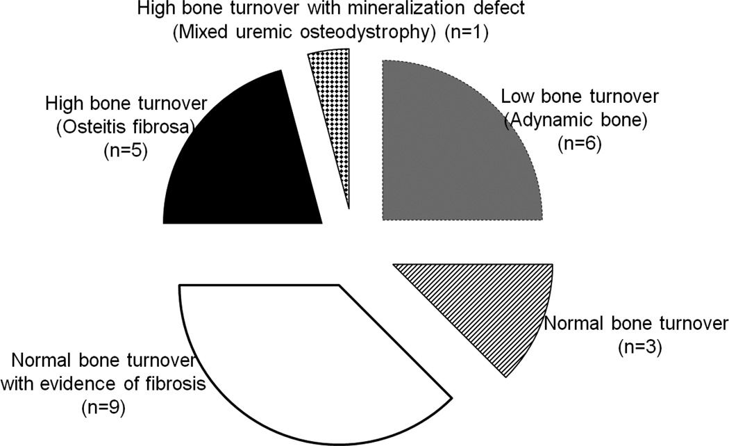
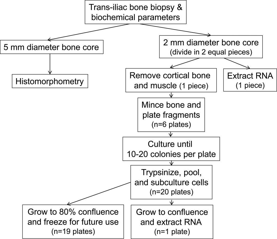
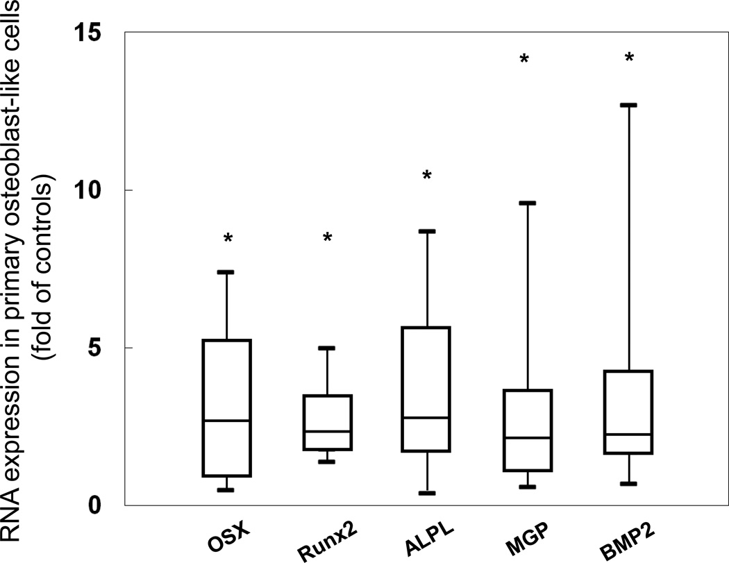
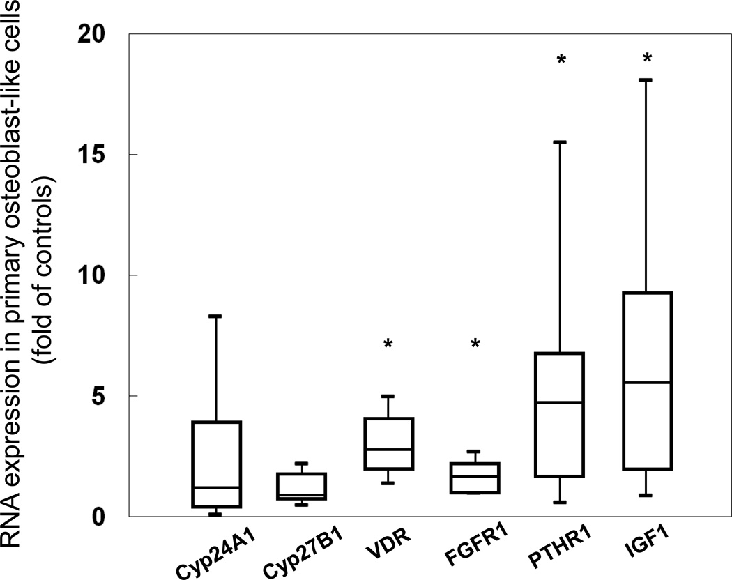
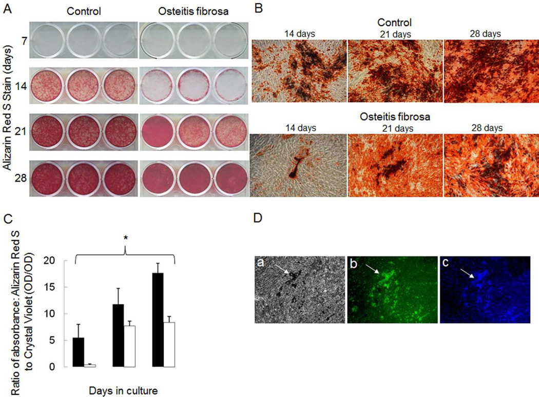
Osteocytes regulate bone turnover and mineralization in chronic kidney disease. As osteocytes are derived from osteoblasts, alterations in osteoblast function may regulate osteoblast maturation, osteocytic transition, bone turnover, and skeletal mineralization. Thus, primary osteoblast-like cells were cultured from bone chips obtained from 24 pediatric ESKD patients. RNA expression in cultured cells was compared with RNA expression in cells from healthy individuals, to RNA expression in the bone core itself, and to parameters of bone histomorphometry. Proliferation and mineralization rates of patient cells were compared with rates in healthy control cells. Associations were observed between bone osteoid accumulation, as assessed by bone histomorphometry, and bone core RNA expression of osterix, matrix gla protein, parathyroid hormone receptor 1, and RANKL. Gene expression of osteoblast markers was increased in cells from ESKD patients and signaling genes including Cyp24A1, Cyp27B1, VDR, and NHERF1 correlated between cells and bone cores. Cells from patients with high turnover renal osteodystrophy proliferated more rapidly and mineralized more slowly than did cells from healthy controls. Thus, primary osteoblasts obtained from patients with ESKD retain changes in gene expression ex vivo that are also observed in bone core specimens. Evaluation of these cells in vitro may provide further insights into the abnormal bone biology that persists, despite current therapies, in patients with ESKD.
骨细胞在慢性肾脏病中调节骨转换和矿化。由于骨细胞来源于成骨细胞,成骨细胞功能的改变可能会调节成骨细胞成熟、骨细胞转变、骨转换和骨骼矿化。因此,从24例儿科终末期肾病(ESKD)患者获取的骨碎片中培养出原代成骨样细胞。将培养细胞中的RNA表达与健康个体细胞中的RNA表达、骨核心本身的RNA表达以及骨组织形态计量学参数进行比较。将患者细胞的增殖和矿化率与健康对照细胞的率进行比较。通过骨组织形态计量学评估的骨类骨质积聚与骨核心中osterix、基质γ-羧基谷氨酸蛋白、甲状旁腺激素受体1和核因子κB受体活化因子配体(RANKL)的RNA表达之间存在相关性。ESKD患者细胞中成骨细胞标志物的基因表达增加,并且包括细胞色素P450 24A1(Cyp24A1)、细胞色素P450 27B1(Cyp27B1)、维生素D受体(VDR)和钠/氢交换调节因子1(NHERF1)在内的信号基因在细胞和骨核心之间具有相关性。高转换型肾性骨营养不良患者的细胞比健康对照者的细胞增殖更快但矿化更慢。因此,从ESKD患者获得的原代成骨细胞在体外保留了在骨核心标本中也观察到的基因表达变化。对这些细胞进行体外评估可能会进一步深入了解ESKD患者尽管目前有治疗方法但仍持续存在的异常骨生物学情况。