Graduate Program in Neuroscience, Department of Neurobiology and Behavior, University of Washington Seattle, WA, USA ; Center for Sensorimotor Neural Engineering, University of Washington Seattle, WA, USA ; Department of Computer Science and Engineering, University of Washington Seattle, WA, USA.
Department of Radiology, University of Washington Seattle, WA, USA.
Front Hum Neurosci. 2014 Oct 16;8:817. doi: 10.3389/fnhum.2014.00817. eCollection 2014.
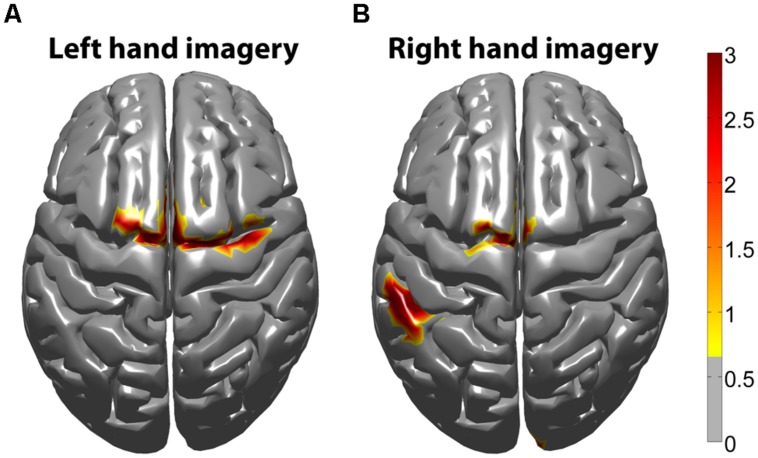
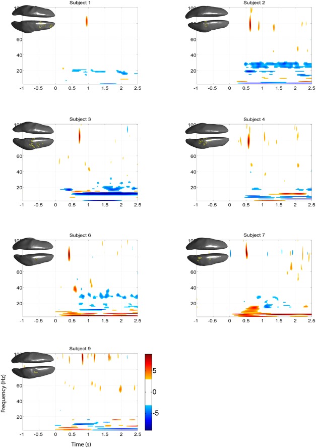
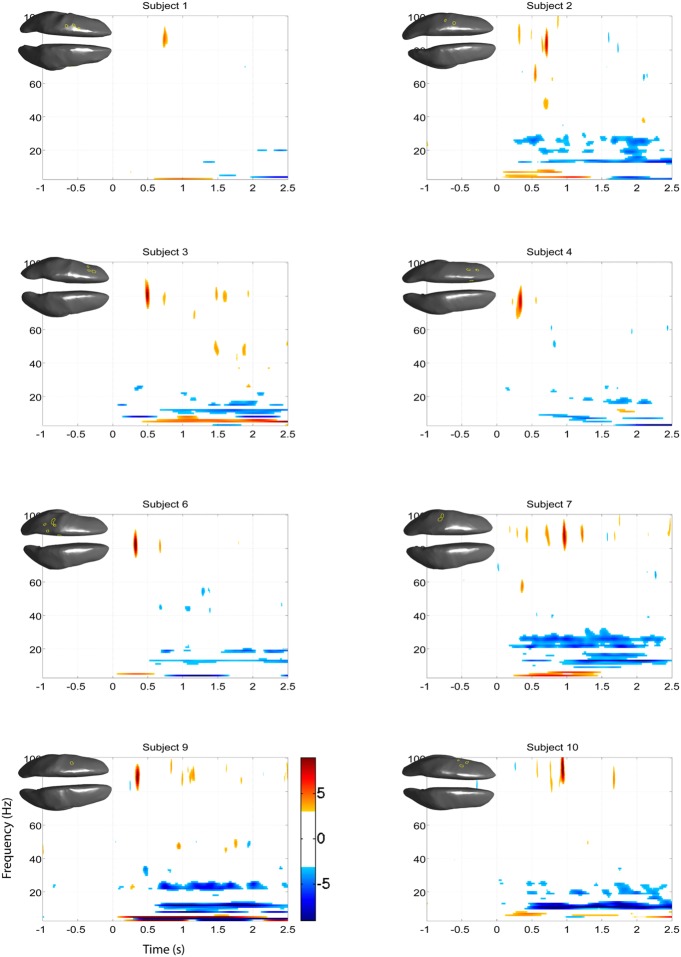
High gamma oscillations (70-150 Hz; HG) are rapidly evolving, spatially localized neurophysiological signals that are believed to be the best representative signature of engaged neural populations. The HG band has been best characterized from invasive electrophysiological approaches such as electrocorticography because of the increased signal-to-noise ratio that results when by-passing the scalp and skull. Despite the recent observation that HG activity can be detected non-invasively by electroencephalography (EEG), it is unclear to what extent EEG can accurately resolve the spatial distribution of HG signals during active task engagement. We have overcome some of the limitations inherent to acquiring HG signals across the scalp by utilizing individual head anatomy in combination with an inverse modeling method. We applied a linearly constrained minimum variance (LCMV) beamformer method on EEG data during a motor imagery paradigm to extract a time-frequency spectrogram at every voxel location on the cortex. To confirm spatially distributed patterns of HG responses, we contrasted overlapping maps of the EEG HG signal with blood oxygen level dependence (BOLD) functional magnetic resonance imaging (fMRI) data acquired from the same set of neurologically normal subjects during a separate session. We show that scalp-based HG band activity detected by EEG during motor imagery spatially co-localizes with BOLD fMRI data. Taken together, these results suggest that EEG can accurately resolve spatially specific estimates of local cortical high frequency signals, potentially opening an avenue for non-invasive measurement of HG potentials from diverse sets of neurologically impaired populations for diagnostic and therapeutic purposes.
高伽马振荡(70-150Hz;HG)是一种快速演变的、空间局部化的神经生理信号,被认为是活跃神经群体的最佳代表性特征。由于绕过头皮和颅骨会导致信噪比增加,因此 HG 带已通过侵入性电生理方法(如皮层电图)得到了最佳描述。尽管最近观察到,通过脑电图(EEG)可以非侵入性地检测到 HG 活动,但尚不清楚 EEG 在多大程度上可以准确解析主动任务参与期间 HG 信号的空间分布。我们通过利用个体头部解剖结构结合逆模型方法,克服了在头皮上获取 HG 信号时固有的一些限制。我们在运动想象范式期间的 EEG 数据上应用了线性约束最小方差(LCMV)波束形成器方法,以在皮层上的每个体素位置提取时频频谱图。为了确认 HG 反应的空间分布模式,我们将 EEG HG 信号的重叠图与来自同一组神经正常受试者的血氧水平依赖(BOLD)功能磁共振成像(fMRI)数据进行对比,这些数据是在单独的会议期间获取的。我们表明,运动想象期间 EEG 检测到的头皮 HG 带活动与 BOLD fMRI 数据在空间上是共定位的。总之,这些结果表明,EEG 可以准确解析局部皮质高频信号的空间特异性估计,这可能为从各种神经损伤人群中无创测量 HG 潜力开辟了一条途径,用于诊断和治疗目的。