• 文献检索
  • 文档翻译
  • 深度研究
  • 学术资讯
  • Suppr Zotero 插件Zotero 插件
  • 邀请有礼
  • 套餐&价格
  • 历史记录
应用&插件
Suppr Zotero 插件Zotero 插件浏览器插件Mac 客户端Windows 客户端微信小程序
定价
高级版会员购买积分包购买API积分包
服务
文献检索文档翻译深度研究API 文档MCP 服务
关于我们
关于 Suppr公司介绍联系我们用户协议隐私条款
关注我们

Suppr 超能文献

核心技术专利:CN118964589B侵权必究
粤ICP备2023148730 号-1Suppr @ 2026

文献检索

告别复杂PubMed语法,用中文像聊天一样搜索,搜遍4000万医学文献。AI智能推荐,让科研检索更轻松。

立即免费搜索

文件翻译

保留排版,准确专业,支持PDF/Word/PPT等文件格式,支持 12+语言互译。

免费翻译文档

深度研究

AI帮你快速写综述,25分钟生成高质量综述,智能提取关键信息,辅助科研写作。

立即免费体验

三维组织学:工具及其在兔心细胞类型分布定量评估中的应用。

Three-dimensional histology: tools and application to quantitative assessment of cell-type distribution in rabbit heart.

机构信息

Department of Physiology, Anatomy and Genetics, University of Oxford, Oxford OX1 3PT, UK.

Department of Physics, University of Oxford, Oxford OX1 3RH, UK.

出版信息

Europace. 2014 Nov;16 Suppl 4(Suppl 4):iv86-iv95. doi: 10.1093/europace/euu234.

DOI:10.1093/europace/euu234
PMID:25362175
原文链接:https://pmc.ncbi.nlm.nih.gov/articles/PMC4217519/
Abstract

AIMS

Cardiac histo-anatomical organization is a major determinant of function. Changes in tissue structure are a relevant factor in normal and disease development, and form targets of therapeutic interventions. The purpose of this study was to test tools aimed to allow quantitative assessment of cell-type distribution from large histology and magnetic resonance imaging- (MRI) based datasets.

METHODS AND RESULTS

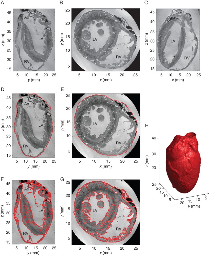
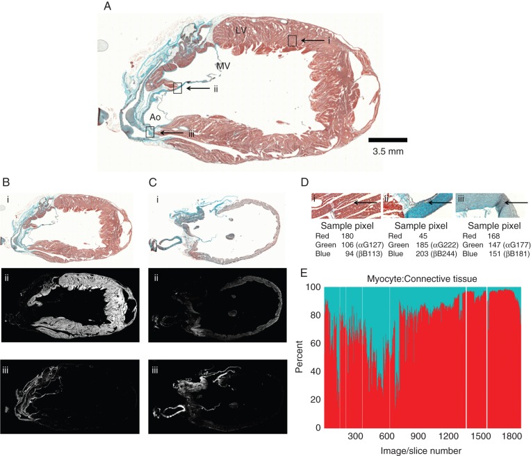
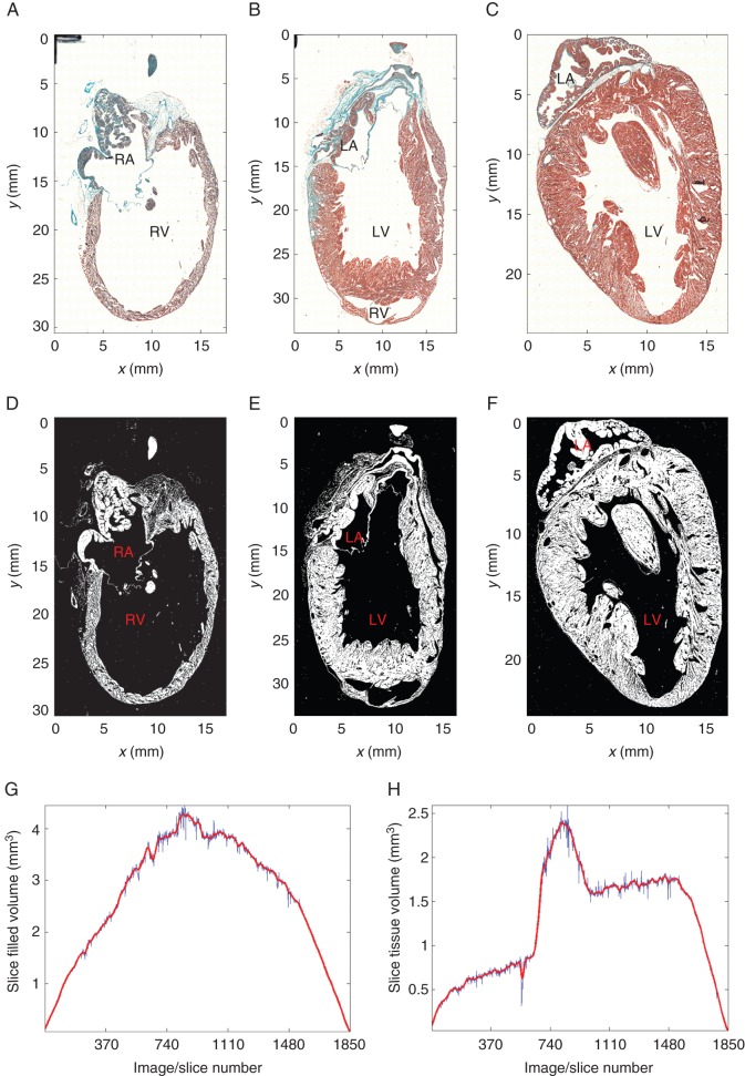
Rabbit heart fixation during cardioplegic arrest and MRI were followed by serial sectioning of the whole heart and light-microscopic imaging of trichrome-stained tissue. Segmentation techniques developed specifically for this project were applied to segment myocardial tissue in the MRI and histology datasets. In addition, histology slices were segmented into myocytes, connective tissue, and undefined. A bounding surface, containing the whole heart, was established for both MRI and histology. Volumes contained in the bounding surface (called 'anatomical volume'), as well as that identified as containing any of the above tissue categories (called 'morphological volume'), were calculated. The anatomical volume was 7.8 cm(3) in MRI, and this reduced to 4.9 cm(3) after histological processing, representing an 'anatomical' shrinkage by 37.2%. The morphological volume decreased by 48% between MRI and histology, highlighting the presence of additional tissue-level shrinkage (e.g. an increase in interstitial cleft space). The ratio of pixels classified as containing myocytes to pixels identified as non-myocytes was roughly 6:1 (61.6 vs. 9.8%; the remaining fraction of 28.6% was 'undefined').

CONCLUSION

Qualitative and quantitative differentiation between myocytes and connective tissue, using state-of-the-art high-resolution serial histology techniques, allows identification of cell-type distribution in whole-heart datasets. Comparison with MRI illustrates a pronounced reduction in anatomical and morphological volumes during histology processing.

摘要

目的

心脏组织解剖结构是功能的主要决定因素。组织结构的变化是正常和疾病发展的一个相关因素,并形成治疗干预的靶点。本研究的目的是测试旨在允许从大型组织学和磁共振成像(MRI)数据集定量评估细胞类型分布的工具。

方法和结果

在心脏停搏期间对兔心进行固定,并进行 MRI 检查,然后对整个心脏进行连续切片,并对三染色组织进行光学显微镜成像。专门为此项目开发的分割技术应用于分割 MRI 和组织学数据集中心肌组织。此外,将组织学切片分割为心肌细胞、结缔组织和未定义组织。为 MRI 和组织学建立了包含整个心脏的边界表面。边界表面内包含的体积(称为“解剖体积”)以及被识别为包含上述任何组织类别的体积(称为“形态体积”)被计算出来。MRI 中的解剖体积为 7.8cm3,组织学处理后减少到 4.9cm3,代表 37.2%的“解剖”收缩。MRI 和组织学之间形态体积减少了 48%,突出了组织水平收缩的存在(例如,间质裂隙空间增加)。分类为包含心肌细胞的像素与被识别为非心肌细胞的像素的比例大致为 6:1(61.6%对 9.8%;其余 28.6%为“未定义”)。

结论

使用最先进的高分辨率连续组织学技术对心肌细胞和结缔组织进行定性和定量区分,允许在整个心脏数据集上识别细胞类型分布。与 MRI 的比较表明,在组织学处理过程中解剖学和形态学体积明显减少。

https://cdn.ncbi.nlm.nih.gov/pmc/blobs/5cc6/4217519/b9bde362595d/euu23403.jpg
https://cdn.ncbi.nlm.nih.gov/pmc/blobs/5cc6/4217519/3d5920ad9c4f/euu23401.jpg
https://cdn.ncbi.nlm.nih.gov/pmc/blobs/5cc6/4217519/514a17a9e67c/euu23402.jpg
https://cdn.ncbi.nlm.nih.gov/pmc/blobs/5cc6/4217519/b9bde362595d/euu23403.jpg
https://cdn.ncbi.nlm.nih.gov/pmc/blobs/5cc6/4217519/3d5920ad9c4f/euu23401.jpg
https://cdn.ncbi.nlm.nih.gov/pmc/blobs/5cc6/4217519/514a17a9e67c/euu23402.jpg
https://cdn.ncbi.nlm.nih.gov/pmc/blobs/5cc6/4217519/b9bde362595d/euu23403.jpg

相似文献

1
Three-dimensional histology: tools and application to quantitative assessment of cell-type distribution in rabbit heart.三维组织学:工具及其在兔心细胞类型分布定量评估中的应用。
Europace. 2014 Nov;16 Suppl 4(Suppl 4):iv86-iv95. doi: 10.1093/europace/euu234.
2
Whole heart detailed and quantitative anatomy, myofibre structure and vasculature from X-ray phase-contrast synchrotron radiation-based micro computed tomography.基于同步辐射 X 射线相衬的微计算机断层扫描的全心详细定量解剖、肌纤维结构和脉管系统。
Eur Heart J Cardiovasc Imaging. 2017 Jul 1;18(7):732-741. doi: 10.1093/ehjci/jew314.
3
Magnetic resonance imaging (MRI) for the assessment of myocardial viability: an evidence-based analysis.用于评估心肌存活性的磁共振成像(MRI):一项基于证据的分析。
Ont Health Technol Assess Ser. 2010;10(15):1-45. Epub 2010 Jul 1.
4
2D and 3D MALDI-imaging: conceptual strategies for visualization and data mining.二维和三维基质辅助激光解吸电离成像:可视化与数据挖掘的概念策略
Biochim Biophys Acta. 2014 Jan;1844(1 Pt A):117-37. doi: 10.1016/j.bbapap.2013.01.040. Epub 2013 Mar 4.
5
[Myocardial microcirculation in humans--new approaches using MRI].[人类心肌微循环——磁共振成像的新方法]
Herz. 2003 Mar;28(2):74-81. doi: 10.1007/s00059-003-2451-6.
6
Visualization and quantification of whole rat heart laminar structure using high-spatial resolution contrast-enhanced MRI.利用高空间分辨率对比增强 MRI 可视化和量化全鼠心脏层状结构。
Am J Physiol Heart Circ Physiol. 2012 Jan 1;302(1):H287-98. doi: 10.1152/ajpheart.00824.2011. Epub 2011 Oct 21.
7
Sub-micrometer anatomical models of the sarcolemma of cardiac myocytes based on confocal imaging.基于共聚焦成像的心肌细胞肌膜亚微米级解剖模型。
Pac Symp Biocomput. 2008:390-401.
8
Positron emission tomography for the assessment of myocardial viability: an evidence-based analysis.用于评估心肌活力的正电子发射断层扫描:基于证据的分析。
Ont Health Technol Assess Ser. 2010;10(16):1-80. Epub 2010 Jul 1.
9
Three-dimensional left ventricular segmentation from magnetic resonance imaging for patient-specific modelling purposes.基于磁共振成像的三维左心室分割用于患者特异性建模。
Europace. 2014 Nov;16 Suppl 4(Suppl 4):iv96-iv101. doi: 10.1093/europace/euu232.
10
The Visible Heart® project and free-access website 'Atlas of Human Cardiac Anatomy'.可视心脏®项目及免费访问网站“人体心脏解剖图谱”
Europace. 2016 Dec;18(suppl 4):iv163-iv172. doi: 10.1093/europace/euw359.

引用本文的文献

1
Full-Field Assessment of Geometry and Collagenous Architecture of Porcine Valve Leaflets via Laser Micrometry and Quantitative Polarized Light Imaging.通过激光测微术和定量偏振光成像对猪瓣膜小叶的几何形状和胶原结构进行全场评估。
J Biophotonics. 2025 Jun;18(6):e202400511. doi: 10.1002/jbio.202400511. Epub 2025 Mar 6.
2
A groupwise registration and tractography framework for cardiac myofiber architecture description by diffusion MRI: An application to the ventricular junctions.基于扩散 MRI 的心肌纤维结构描述的分组配准和束追踪框架:在心室交界处的应用。
PLoS One. 2022 Jul 18;17(7):e0271279. doi: 10.1371/journal.pone.0271279. eCollection 2022.
3

本文引用的文献

1
Cardiac fibroblasts: the good, the bad, the ugly, the beautiful.心脏成纤维细胞:有益的、有害的、丑陋的、美好的。
J Mol Cell Cardiol. 2014 May;70:1. doi: 10.1016/j.yjmcc.2014.03.009. Epub 2014 Mar 17.
2
Fibroblast-myocyte electrotonic coupling: does it occur in native cardiac tissue?成纤维细胞-心肌细胞电偶联:在天然心脏组织中是否存在?
J Mol Cell Cardiol. 2014 May;70(100):37-46. doi: 10.1016/j.yjmcc.2013.12.024. Epub 2014 Jan 8.
3
Fibroblasts in myocardial infarction: a role in inflammation and repair.心肌梗死后的成纤维细胞:在炎症和修复中的作用。
Impact of intraventricular septal fiber orientation on cardiac electromechanical function.
室间隔纤维方向对心机电功能的影响。
Am J Physiol Heart Circ Physiol. 2022 Jun 1;322(6):H936-H952. doi: 10.1152/ajpheart.00050.2022. Epub 2022 Mar 18.
4
Transformation diffusion reconstruction of three-dimensional histology volumes from two-dimensional image stacks.从二维图像堆叠中重建三维组织学体积的变换扩散。
Med Image Anal. 2017 May;38:184-204. doi: 10.1016/j.media.2017.03.004. Epub 2017 Mar 23.
5
Resolving Fine Cardiac Structures in Rats with High-Resolution Diffusion Tensor Imaging.利用高分辨率扩散张量成像解析大鼠精细心脏结构
Sci Rep. 2016 Jul 28;6:30573. doi: 10.1038/srep30573.
6
Mapping cardiac microstructure of rabbit heart in different mechanical states by high resolution diffusion tensor imaging: A proof-of-principle study.通过高分辨率扩散张量成像绘制不同力学状态下兔心脏的心脏微观结构:一项原理验证研究。
Prog Biophys Mol Biol. 2016 Jul;121(2):85-96. doi: 10.1016/j.pbiomolbio.2016.06.001. Epub 2016 Jun 16.
7
Human-based approaches to pharmacology and cardiology: an interdisciplinary and intersectorial workshop.基于人体的药理学与心脏病学方法:一场跨学科和跨部门研讨会。
Europace. 2016 Sep;18(9):1287-98. doi: 10.1093/europace/euv320. Epub 2015 Nov 29.
8
Myocardial interstitial remodelling in non-ischaemic dilated cardiomyopathy: insights from cardiovascular magnetic resonance.非缺血性扩张型心肌病中心肌间质重塑:心血管磁共振成像的见解
Heart Fail Rev. 2015 Nov;20(6):731-49. doi: 10.1007/s10741-015-9509-4.
9
Three-dimensional cardiac computational modelling: methods, features and applications.三维心脏计算建模:方法、特点与应用
Biomed Eng Online. 2015 Apr 17;14:35. doi: 10.1186/s12938-015-0033-5.
10
Interrogation of living myocardium in multiple static deformation states with diffusion tensor and diffusion spectrum imaging.利用扩散张量成像和扩散谱成像对处于多种静态变形状态的活体心肌进行检测。
Prog Biophys Mol Biol. 2014 Aug;115(2-3):213-25. doi: 10.1016/j.pbiomolbio.2014.08.002. Epub 2014 Aug 10.
J Mol Cell Cardiol. 2014 May;70:74-82. doi: 10.1016/j.yjmcc.2013.11.015. Epub 2013 Dec 7.
4
Origin, development, and differentiation of cardiac fibroblasts.心脏成纤维细胞的起源、发展和分化。
J Mol Cell Cardiol. 2014 May;70:2-8. doi: 10.1016/j.yjmcc.2013.11.003. Epub 2013 Nov 11.
5
Validation of contrast-enhanced magnetic resonance imaging to monitor regenerative efficacy after cell therapy in a porcine model of convalescent myocardial infarction.在猪心肌梗死后恢复期细胞治疗模型中,使用对比增强磁共振成像监测再生疗效的验证。
Circulation. 2013 Dec 24;128(25):2764-75. doi: 10.1161/CIRCULATIONAHA.113.002863. Epub 2013 Sep 23.
6
Patient-Specific Models of Cardiac Biomechanics.心脏生物力学的个性化模型
J Comput Phys. 2013 Jul 1;244:4-21. doi: 10.1016/j.jcp.2012.09.015.
7
Combining wet and dry research: experience with model development for cardiac mechano-electric structure-function studies.结合湿实验和干实验:心脏力学-电学结构-功能研究模型开发经验。
Cardiovasc Res. 2013 Mar 15;97(4):601-11. doi: 10.1093/cvr/cvt003. Epub 2013 Jan 17.
8
Computational cardiology: the heart of the matter.计算心脏病学:问题的核心
ISRN Cardiol. 2012;2012:269680. doi: 10.5402/2012/269680. Epub 2012 Nov 14.
9
Histo-anatomical structure of the living isolated rat heart in two contraction states assessed by diffusion tensor MRI.应用扩散张量 MRI 评估两种收缩状态下活体分离大鼠心脏的组织解剖结构。
Prog Biophys Mol Biol. 2012 Oct-Nov;110(2-3):319-30. doi: 10.1016/j.pbiomolbio.2012.07.014. Epub 2012 Aug 7.
10
Methodology for patient-specific modeling of atrial fibrosis as a substrate for atrial fibrillation.将心房纤维化作为心房颤动基质进行患者特异性建模的方法。
J Electrocardiol. 2012 Nov-Dec;45(6):640-5. doi: 10.1016/j.jelectrocard.2012.08.005. Epub 2012 Sep 19.